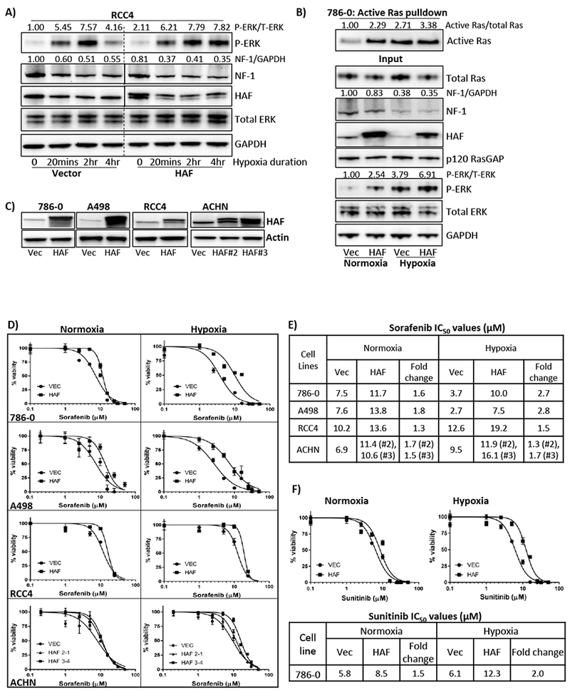
Figure 4: HAF promotes increased Ras-ERK pathway activation and is associated with resistance to sorafenib.

A) Impact of increasing duration of hypoxia on levels of neurofibromin and p-ERK in vector or HAF overexpressing RCC4 cells. Normalized densitometric values for p-ERK and neurofibromin relative to total ERK and GAPDH respectively are indicated. B) Results of active Ras pulldown assay in 786-0 cells stably expressing empty vector or HAF exposed to normoxia or hypoxia for 2 hours. Normalized densitometric values for active GTP-bound Ras relative to total Ras are indicated. C) Western blot showing HAF levels in a panel of stably generated vector or HAF overexpressing CCRCC cell lines. D-F) XTT viability assays of indicated cell lines after 72 hours’ continuous treatment with sorafenib (D), or sunitinib (F) with calculated IC50 values in (E-F). Data shown are representative of at least two independent experiments.
