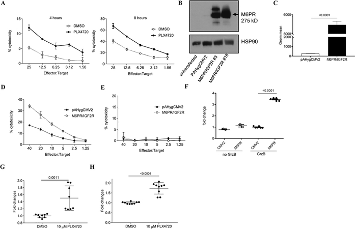
Figure 3. M6PR up-regulation on the cell surface sensitizes WM35 cells to the cytotoxic effect of TIL.
A. After pretreatment of target cells (WM35) with PLX4720 O/N, 4–8 hours Cr release assay was performed in triplicate. Cells were labeled with 51Cr and cultured with effector cells (HLA-A2+ TIL) at the indicated ratios. Appropriate maximum and minimum release controls were determined in each experiment. Representative results of 3 different experiments are shown. B. Analysis of total protein from control (pAHygCMV2) or M6PR over-expressing (M6PR/IGF2R) WM35 cells. Lysates were probed with M6PR and HSP90 specific antibodies. C. Cell surface levels of M6PR was determined by flow cytometry in control (WM35-pAHygCMV2) and M6PR-overexpressing cells (WM35-IGF2R/M6PR). Bars represent standard deviation (SD). Statistical analysis was done using unpaired two-tailed Student’s t test. D, E. 6 hours 51Cr experiment was performed in triplicates. As target cells, 51Cr labeled WM35-pAHygCMV2 (control) and WM35- IGF2R/M6PR were used and cultured with HLA-matching TIL (D) or healthy donor derived CD8+ T cells (E). F. Cells were incubated with inactive GrzB for 1 hr and intracellular GrzB level was measured by flow cytometry. Mean and SD of individual experiments are shown (n=6). P values were calculated in two-sided Student’s t-test. G, H. WM35 cells were treated with DMSO or PLX4720 O/N and cell surface levels of M6PR was detected by flow cytometry (G). H. GrzB uptake by WM35 cells were detected by intracellular GrzB staining using GrzB specific mouse anti-human antibody. Geometric mean was calculated and all results were normalized to control (DMSO-treated cells). Individual results and SD are shown. P values are shown in unpaired two-tailed Student’s t test.

