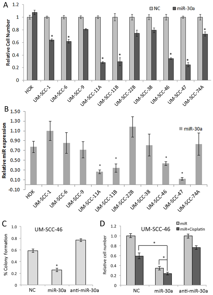
Fig. 3. A miR-30a-5p mimic inhibited HNSCC cell proliferation, colony formation, and enhanced cisplatin sensitivity in vitro.
(A) Proliferation measured by XTT assay in 6 replicates on day 5 following transfection with control or miR-30a-5p mimic across primary Human Oral kerytinocytes (HOK) and ten HNSCC cell lines. (B) Basal level of miR-30a-5p expression measured by qRT-PCR in HOK and ten HNSCC cell lines in log-growth phase. Relative miR-30a-5p expression was normalized to the mean expression of the cell lines. * denotes p< 0.05 by a Student’s t-test compared to HOK cells. (C) Colony formation assay of UM-SCC-46 cells was performed following 48 h transfection with miR-30a-5p or anti-miR30a-5p oligonucleotides. Colonies were counted in three wells and repeated in three independent experiments. (D) UM-SCC-46 cells were transfected with miR-30a-5p mimic for 48 hrs, and treated with 2µM cisplatin for 3 h and then washed with warm media and cell density was measured by XTT assay 72 h after cisplatin treatment. Values represent the mean of at least three experiments ±SEM, * denotes p< 0.05 by a Student’s t-test.

