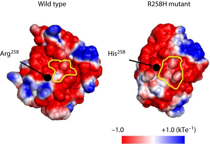
Figure 1.

Electrostatic surface potential of wild‐type and R258H mutant forms of hepatocyte nuclear factor 4α. Potentials from negative to positive are shown as red to blue, respectively. Dimer interfaces are outlined in yellow. [Colour figure can be viewed at wileyonlinelibrary.com]
