
Figure 8.
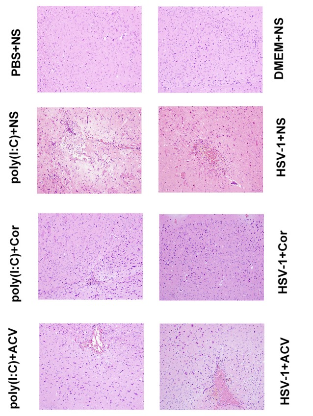
Corilagin induces histopathological changes in the brain of mice with encephalitis. Histopathological changes in the right temporal lobe brain tissue were observed by hematoxylin-eosin (HE) staining (×200). DMEM (20 μL), phosphate-buffered saline (PBS; 20 μL), poly(I:C; 5 mg/kg, 20 μL), or HSV-1 (10−4/20 μL) were injected into the intracalvarium at the midpoint of the line from the right canthus to the external auditory canal. One hour after the model was established, five mice in each group were given normal saline (NS), corilagin 40 mg/kg, or ACV 350 mg/kg daily intragastrically. The brain tissues were taken for HE staining.