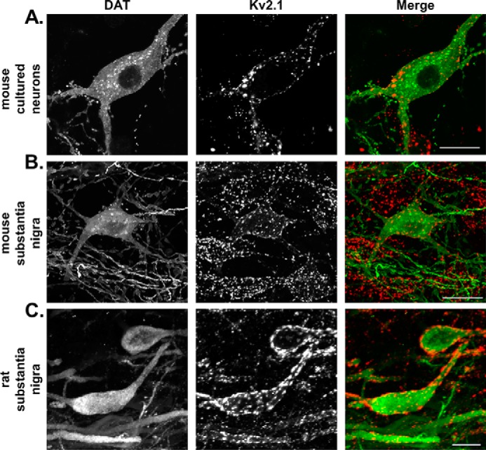
Figure 1.

Kv2.1 is expressed at the soma and proximal processes of murine dopamine neurons. Double-immunofluorescence labeling is shown of DAT (leftmost column, green in merge) and Kv2.1 (middle column, red in merge) in murine dopamine neurons. All images are 2D maximum intensity projections of 3D reconstructed confocal z-stack. In mouse cultured neurons (A) and labeled mouse (B) and rat (C) brain slices, DAT labeling is diffuse along the entirety of the soma and proximal processes, whereas Kv2.1 is localized exclusively to large clusters. (Scale, 10 μm.)
