Figure 1.
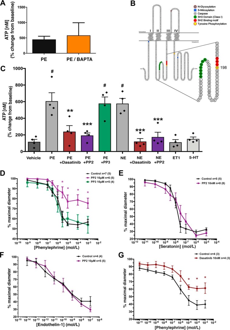
SRC family kinases regulate phenylephrine-induced pannexin 1 channel function independent of Ca2+. A, PE-stimulated ATP release from intact TDAs treated with the intracellular calcium-chelating agent BAPTA-AM. n = 4 TDAs (from four mice). Data are presented as percentage increase from the unstimulated condition. Student's t test was performed to test for significance. B, mouse pannexin 1 membrane topology denoting SH2 linear motif (red) containing the putative intracellular loop tyrosine 198 (yellow) SRC phosphorylation site and the upstream proline-rich SH3-binding domain (green). C, ex vivo ATP release from TDA stimulated with PE (20 μm) and norepinephrine (10 μm) in the presence and absence of the SFK inhibitors PP2 (10 μm), dasatinib (10 nm), and negative control PP3 (10 μm). The nonadrenergic vasoconstrictors 5-HT (100 nm) and ET-1 (10 nm) were assessed. n = 4 TDA (from four mice) per group. One-way ANOVA was performed for statistical significance. #, significant increase from vehicle; p < 0.5. Asterisks, significant reduction from PE-stimulated; *, p < 0.05; **, p < 0.01; ***, p < 0.001. D–G, contractile responses to increasing concentration of adrenergic agonists (PE or norepinephrine) in the presence of SFK inhibitors PP2 (10 μm; purple line), inactive control PP3 (10 μm; green line), and dasatinib (10 nm; red line). n = 4–8 TDA (from 4–8 mice). Data are presented as mean ± S.E. (error bars) and were assessed for significance using two-way ANOVA with Bonferroni post hoc test of multiple comparisons. *, p < 0.05 compared with control response (black line).

