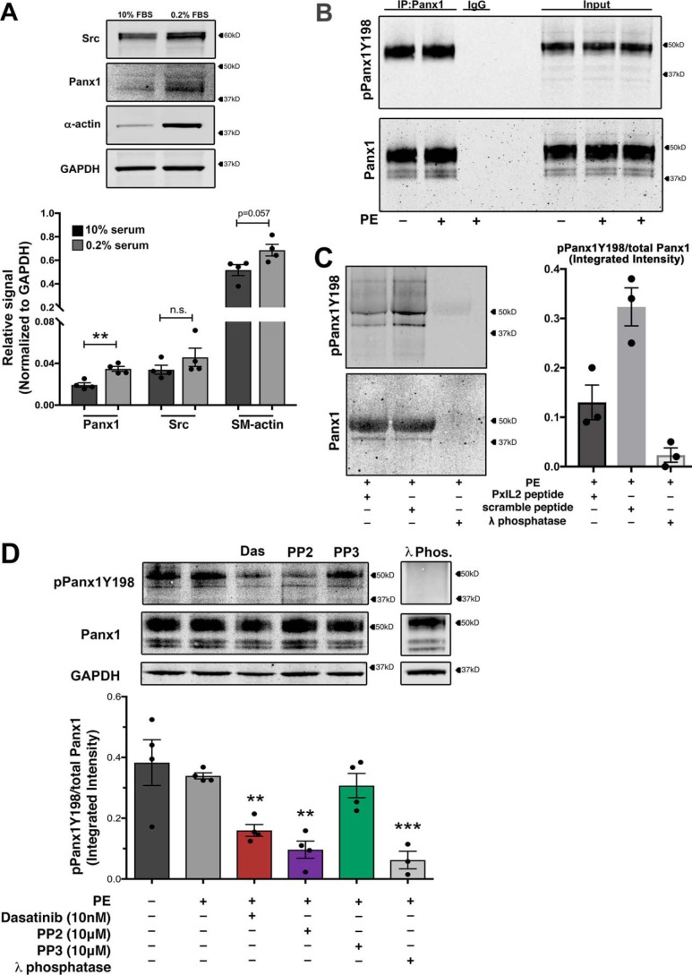
Figure 2.
Pannexin 1 tyrosine 198 is constitutively phosphorylated in vascular smooth muscle cells. A, confirmation of protein expression of PANX1 and SRC kinase in smooth muscle cells following differentiation to a contractile phenotype with 48-h serum starvation (0.2% FBS). Data are presented as mean ± S.E. (error bars), n = 4 independent experiments. Student's t test was performed to test for statistical significance. *, p < 0.05; **, p < 0.01 compared with high FBS level. B, immunoprecipitation of PANX1 from differentiated hCoSMCs stimulated with PE (20 μm) and immunoblotted for pannexin 1 (Tyr198) phosphorylation. C, the PxIL2 peptide was used at 10 mm to inhibit pPANX1Y198 phosphorylation after adrenergic stimulation. λ-Phosphatase was used to eliminate all tyrosine phosphorylation of PANX1. D, representative Western blotting and quantification of phosphorylation status of pannexin 1 Tyr198 in hCoSMCs treated with SFK inhibitors dasatinib (10 nm; red), PP2 (10 μm; purple), and negative control PP3 (10 μm; green). λ-Phosphatase–treated lysates were used as a negative control; n = 6 independent experiments. Data quantification is presented as mean ± S.E. *, p < 0.05; **, p < 0.01; ***, p < 0.001 compared with unstimulated control using one-way ANOVA.

