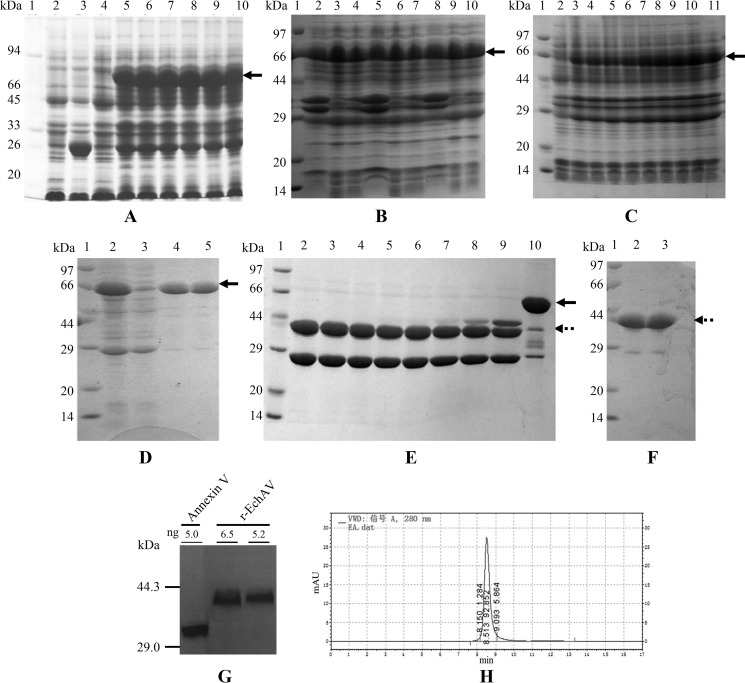
Figure 2.
Optimal conditions for inducing the expression, purification, and determination of r-EchAV. Samples were analyzed by 12% SLS-PAGE followed by Coomassie Blue staining. All samples were boiled for 3 min with 50 mm DTT. Approximately 10–15 μg of protein was loaded on each lane. The reference range of protein molecular mass is 97–14 kDa. The induced expression band of the target protein is indicated by the arrow in A–C. D–F, GST–r-EchAV is shown by the solid arrow; r-EchAV is shown by the dotted arrow. A, identification of different concentrations of IPTG as inducer. Lane 1, molecular mass markers; lane 2, E. coli/pGEX-6P-1, no IPTG addition; lane 3, E. coli/pGEX-6P-1, 100 μm IPTG; lanes 4–10, E. coli/pGEX-6P–r-EchAV, IPTG addition was at different concentrations: 0, 25, 50, 100, 150, 200, and 300 μm, respectively. B, identification under different induction temperatures. Lane 1, molecular mass markers; lanes 2–4, total bacterial protein, supernatant, and inclusion body obtained by induced expression at 37 °C; lanes 5–7, total bacterial protein, supernatant, and inclusion body obtained by induced expression at 30 °C; lanes 8–10, total bacterial protein, supernatant, and inclusion body obtained by induced expression at 25 °C. C, identification after different induction times. Lane 1, molecular mass markers; lanes 2–11, the total bacterial protein after 0 and 5–12, or 20 h of IPTG induced expression. D, purification of induced fusion protein GST–r-EchAV by affinity chromatography. Lane 1, molecular mass markers; lane 2, supernatant after induced expression at 25 °C; lane 3, penetrating solution in chromatography; lanes 4 and 5, eluted GST–r-EchAV. E, cleavage efficiency of GST–r-EchAV by PSP-specific protease digestion. Lane 1, molecular mass markers; lanes 2–9, PSP digestion effect (molar ratio of GST–r-EchAV to PSP enzyme is 800, 400, 300, 200, 100, 50, 10, and 3, respectively); lane 10, fusion protein as a control. F, purified r-EchAV was identified by 12% SLS-PAGE. Lane 1, molecular mass markers; lanes 2 and 3, r-EchAV. G, Western blotting assay of r-EchAV (44.1 kDa) and annexin V (35.7 kDa). H, SEC-HPLC analysis of r-EchAV.

