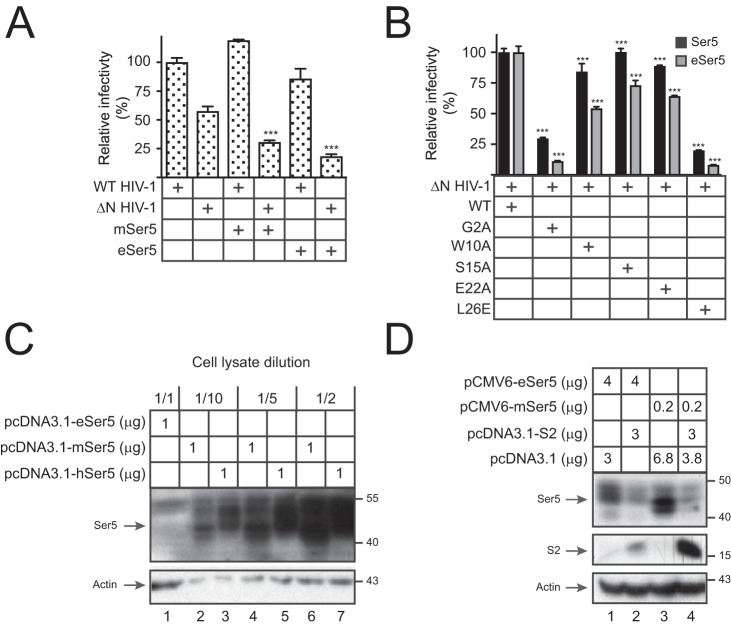
Figure 7.
Analysis of eSer5 antiviral activity and sensitivity to S2. A, WT and ΔN HIV-1 pseudoviruses were produced from 293T cells in the absence or presence of 0.2 μg of pcDNA3.1-mSer5-FLAG or pcDNA3.1-eSer5-FLAG. Viral infectivity was measured and presented as in Fig. 1A. B, ΔN HIV-1 pseudoviruses were produced from 293T cells in the presence of pcDNA3.1-mSer5-FLAG or pcDNA3.1-eSer5-FLAG and the indicated S2 expression vectors. Viral infectivity was measured and presented as in Fig. 1A. C, 293T cells were transfected with 1 μg of pcDNA3.1-eSer5-FLAG, pcDNA3.1-mSer5-FLAG, or pcDNA3.1-hSer5-FLAG. Lysate from cells expressing human and murine Ser5 was diluted as indicated, and Ser5 expressions were compared by Western blotting. D, 293T cells were transfected with 3 μg of pcDNA3.1-S2-HA and 4 μg of pCMV6-eSer5-FLAG or 0.2 μg of pCMV6-mSer5-FLAG. Ser5 and S2 expressions were determined by Western blotting. Error bars in A and B indicate S.E. from three independent experiments. ***, p < 0.001.

