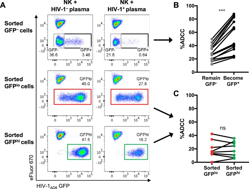
FIG 5.
ADCC against HIV-1AD8-infected CEM cells sorted into GFP–, GFPlo, and GFPhi populations. (A) Representative scatter plots showing elimination of the sorted target cells in the presence of effector NK cells with HIV-1+ plasma compared to NK cells with HIV-1– plasma. Uninfected CEM cells stained with the cell proliferation dye eFluor 670 (eFluor 670+ GFP–) were spiked into each sample at the end of the 5-h ADCC assay incubation to serve as a reference population to calculate percent ADCC of the sorted target cells. (B) ADCC mediated by HIV-1+ plasma (1:1,000; n = 14) against sorted GFP– cells that remained GFP– or became GFP+ after the 5-h assay incubation. (C) ADCC mediated by HIV-1+ plasma (1:1,000; n = 8) against sorted GFPlo and GFPhi cells. Statistical analyses between matched pairs were performed with a Wilcoxon signed-rank test (ns, not significant; ***, P < 0.001).

