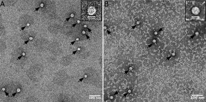
FIG 3.

Electron micrographs of capsid preparations. (A) Capsids that had been extensively purified through method I. (B) Capsids that had been only partially purified. Arrows point to individual assembled capsids. (Insets) Enlarged images of single capsids to better assess size and shape. Scale bars are included.
