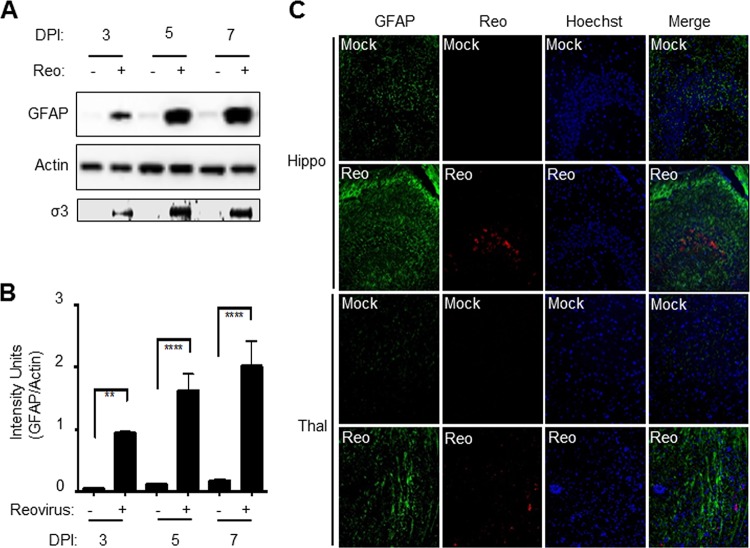
FIG 1.
Reovirus infection of the brain results in increased expression of GFAP. Two-day-old Swiss Webster (SW) mice were infected with reovirus by i.c. inoculation of 100 PFU of reovirus (T3 Dearing/T3D). (A) Western blot analysis (see representative blot) shows increased GFAP levels at 3, 5, and 7 days p.i. (DPI) in reovirus (Reo)-infected brains compared to mock-infected controls. Increased reovirus antigen (σ3) was also seen at 3, 5, and 7 days p.i. Actin levels were used to demonstrate equivalent protein loading between samples. (B) Densitometric analysis of three blots. The graph shows the mean intensity of GFAP bands. Error bars represent the standard errors of the mean (SEM). **, P < 0.01; ****, P < 0.0001. (C) At 8 days p.i., increased GFAP (green) and reovirus σ3 (red) staining in virus-infected brains (Reo) compared to mock-infected controls can be seen in and around the hippocampus (hippo) and thalamus (thal), two areas targeted by reovirus.

