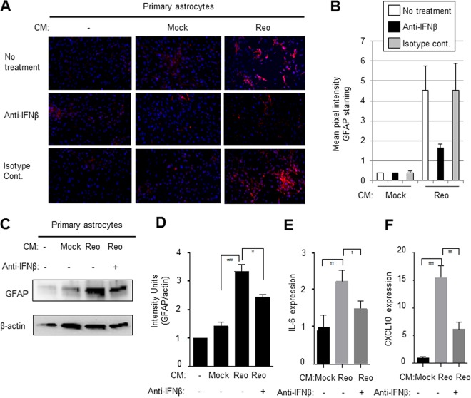
FIG 5.
The ability of supernatants from reovirus-infected BSCs to activate primary astrocytes is blocked with anti-IFN-β antibody. Conditioned medium (CM) was collected from mock-infected and reovirus (Reo)-infected BSCs at 8 days p.i. and was added to primary astrocytes (ratio of reovirus conditioned medium [BSC supernatant] to fresh DMEM = 1/10) in the presence of anti-IFN-β antibodies or isotype-matched controls. Fresh BSC medium was also used as a control. (A) Three days following treatment, the astrocytes were stained with GFAP. Increased expression of GFAP (red) was seen in astrocytes exposed to reovirus-conditioned medium compared to astrocytes exposed to medum from mock-infected BSCs or medium alone. This activation was decreased when the conditioned medium was first treated with anti-IFN-β antibody (10 µg/ml) but remained unchanged when conditioned medium was treated with an isotype-matched antibody (10 µg/ml). (B) A graph shows the mean pixel intensity of images taken from the center of three wells of a chamber slide from astrocytes exposed to media from mock- or virus-infected primary astrocytes. (C to F) Increased expression of GFAP (C and D) and cytokines (E and F) in primary astrocytes treated with conditioned medium from reovirus-infected BSCs compared to astrocytes treated with medium from mock-infected BSCs, as shown by Western blotting (C and D) or RT-PCR analysis (E and F) and was blocked in the presence of anti-IFN-β antibodies. Graphs D to F show the mean expression of GFAP or chemokines/cytokines. Error bars represent the SEM. *, P < 0.05; **, P < 0.01; ***, P < 0.001; ****, P < 0.0001.

