FIG 6.
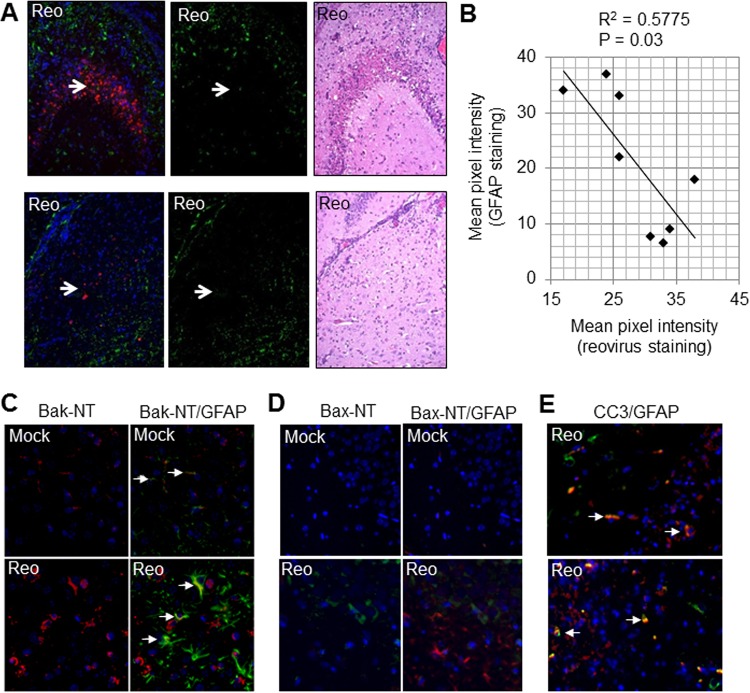
As infection progresses, astrocytes undergo apoptosis. Two-day-old SW mice were infected with reovirus (Reo) as described previously (Fig. 1). At 8 days p.i., mice were sacrificed, and brains were sectioned and prepared for IHC. (A) In areas with high levels of virus antigen (red staining) and injury (H&E staining), GFAP staining (green) is absent (arrows). (B) The red (reovirus antigen) and green (GFAP) pixel intensity for the CA2 region of the hippocampus of eight reovirus-infected mice was quantified using ImageJ. The graph shows high GFAP staining when reovirus antigen is present in relatively small amounts, but that GFAP expression drops off as levels of reovirus antigen increase. (C) Images from the thalamus demonstrating increased Bak-NT staining (red) in reovirus-infected sections compared to mock-infected controls. Bak-NT staining colocalized with GFAP staining (green) in some astrocytes (arrows). (D) Images from the hippocampus demonstrating that Bax-NT staining (green) increased in virus-infected sections compared to mock-infected controls but did not colocalize with GFAP (red). (E) Images from the hippocampus demonstrate that cleaved caspase 3 (CC3, red) colocalizes with GFAP (green) in some astrocytes (arrows). Cells staining for cleaved caspase 3 but not GFAP are likely reovirus-infected neurons.

