FIG 8.
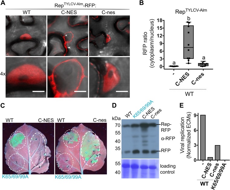
Rep nuclear localization is essential for its DNA replication activity. (A) Subcellular localization of WT RepTYLCV-Alm when an NES or a nonfunctional nes is fused to its C terminus in N. benthamiana, visualized at 3 days after agroinfiltration in cells (top) and their nuclei (bottom). Scale bars represent 5 μm. The arrowhead indicates Rep-RFP in the cytoplasm, and the asterisk indicates the nonfluorescent nucleus. (B) Box plot showing the ratio of RFP fluorescence intensity in the cytoplasm versus nucleus for the images shown in panel A. Ten cells per sample were analyzed under the same conditions as for Fig. 2. (C) UV image of leaves from 2IRGFP N. benthamiana plants that transiently express C-NES/nes fusions of WT Rep (right half of the leaf) and the nontagged Reps as a control (WT and nonfunctional triple K-to-A mutant) (left half). (D) Immunoblot of the total protein extracts from agroinfiltrated leaf areas, revealing the protein levels of WT Rep-RFP and its variants (anti-RFP). To confirm equal protein loading for each sample, the membrane was stained with Coomassie brilliant blue. (E) Quantification of the circular extrachromosomal molecules (ECMs) in the agroinfiltrated 2IRGFP leaf areas using real-time PCR on total DNA extracts.

