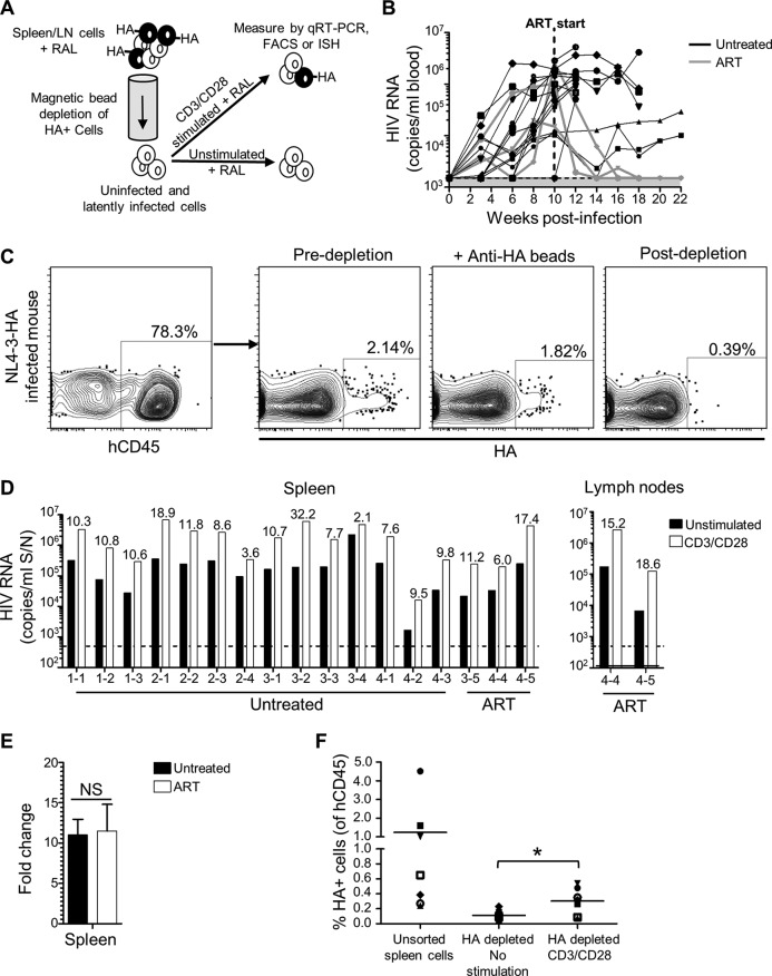
FIG 4.
Detection of latent HIV-1 in humanized mice. (A) Schematic of ex vivo latency reactivation assay. HIV-infected/HA+ cells from disaggregated spleen or lymph node suspensions were removed using anti-HA antibodies and magnetic bead depletion, and the resulting cells were cultured with RAL in either unstimulating conditions or in the presence of anti-CD3/CD28 antibodies. After 2 days, HIV-1 was measured by qRT-PCR of culture supernatants or by FACS or ISH analysis of cells. (B) Seventeen humanized mice from four cohorts were infected with NL4-3-HA and HIV-1 levels in blood monitored over time by qRT-PCR. The shaded area is the LOD, i.e., 1,500 copies/ml. After 10 weeks, three animals were put on oral ART. All mice were necropsied at the indicated time points. Spleens were harvested from all animals, and lymph nodes were also harvested from two of the ART-treated mice. (C) FACS plots showing depletion of HA+ cells by anti-HA antibodies and magnetic beads. (D) HIV-1 RNA levels in culture supernatants from the ex vivo latency assay after 2 days in either unstimulated or CD3/CD28-stimulated cultures. Individual spleen or lymph node samples are shown from indicated mice (mouse IDs from cohorts 1 to 4 are indicated). The dotted line is the LOD, i.e., 500 copies/ml. Numeric values indicate the fold-increase in HIV-1 production from matched stimulated versus unstimulated cultures. (E) Mean fold increase from spleen samples combined for ART-treated versus untreated mice. NS, not significant. (F) HA expression was measured by flow cytometry for human CD45+ cells in bulk unsorted spleen samples at the time of isolation or for the HA-depleted populations after 2 days of culture in either unstimulated or stimulated conditions. Samples are from the seven untreated animals in cohorts 1 and 2. *, P < 0.05.

