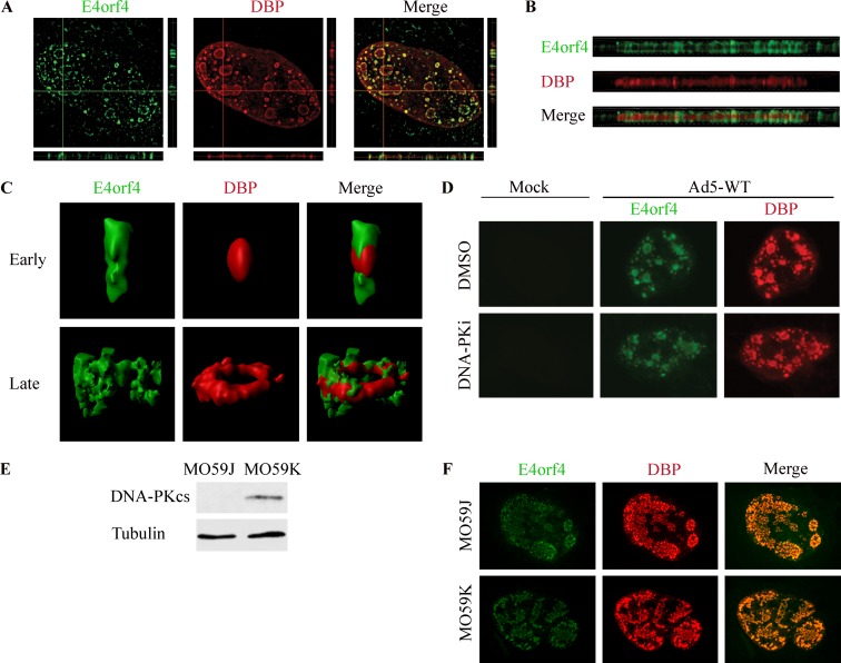
FIG 5.
E4orf4 is recruited to viral RCs in a DNA-PK-independent manner. (A) HeLa cells were infected with WT-Ad5 and were fixed and stained at 24 h p.i. A high-resolution image of a representative infected nucleus is shown with horizontal and vertical orthogonal projections. (B) Enlarged image of a horizontal orthogonal projection from the nucleus presented in panel A. (C) Representative 3D renderings of early and late replication centers prepared with Imaris software. (D) HeLa cells were either mock infected or infected with WT-Ad5 in the presence of the solvent dimethyl sulfoxide (DMSO) or the DNA-PK inhibitor (DNA-PKi) NU7441. Representative confocal images of cells fixed at 24 h p.i. are shown. (E) MO59K and MO59J cell extracts were subjected to Western blot analysis with the indicated antibodies. (F) MO59K and MO59J cells were infected with WT-Ad5, fixed at 24 h p.i., and subjected to confocal microscopy. Representative images are shown.

