
Figure 5.
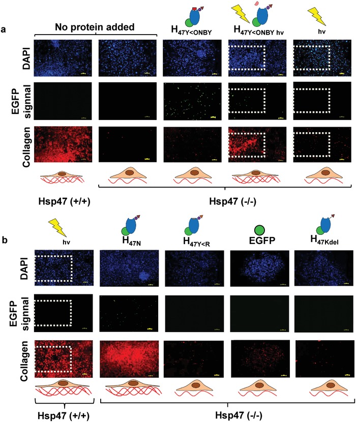
Localized induction of collagen deposition by photoactivation of photoactivatable Hsp47. Immunostaining of MEF Hsp47 +/+ and −/− cultures 24 h after treatment with different Hsp47 variants and controls Col1 antibody staining in red, Hsp47 variants in green (EGFP), and nuclei in blue (DAPI). a) Hsp47 +/+ and −/− having no protein delivery were used as controls. The light exposed areas (1.8 × 1.2 mm2) are highlighted with dotted square. Irradiation wavelength was 405 nm. b) Hsp47 +/+ cells incubated with other inactive mutants did not show enhanced collagen production, whereas cells incubated with H47N showed higher collagen levels. Scale bar: 250 µm.