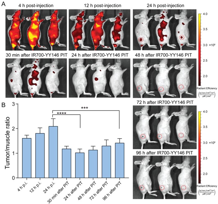
Figure 6.

Serial IR700 fluorescence imaging before and after IR700‐YY146 PIT of small melanomas. A) Fluorescence imaging using an IVIS Spectrum was performed at different time‐points before and after NIR irradiation. Tumors were indicated by red dashed circles. B) The mean fluorescence intensity ratios of tumor to muscle were quantitatively calculated before and after IR700‐YY146 PIT (***p < 0.001, ****p < 0.0001, p = 4 for the group).
