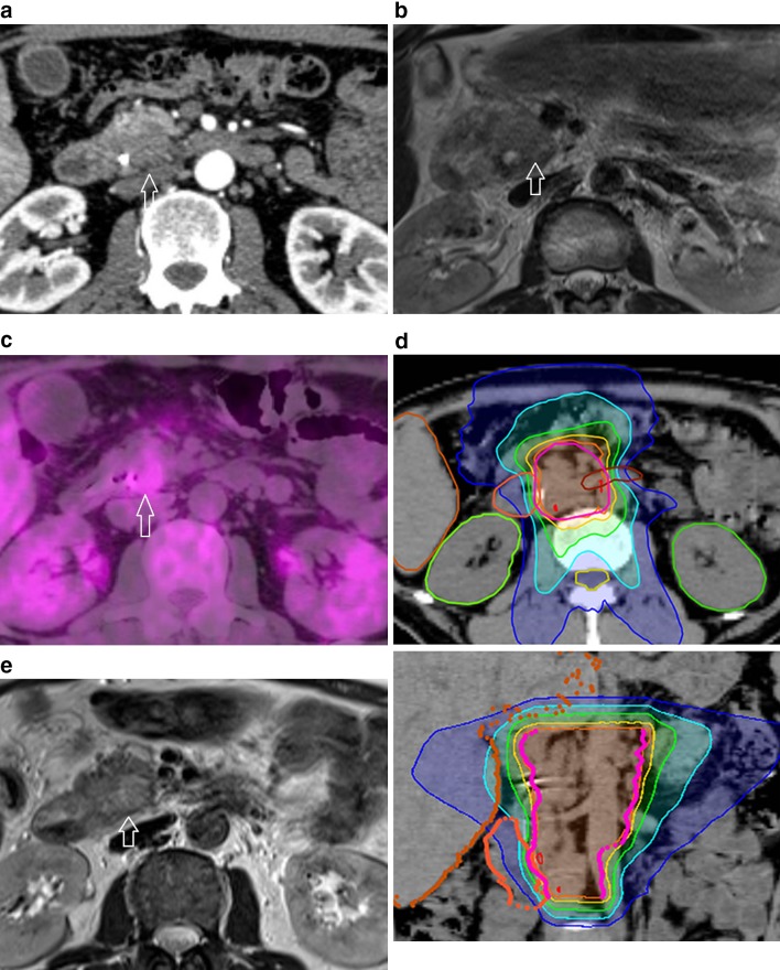
Fig. 1.
a Axial contrast-enhanced CT in the arterial phase shows a hypo-enhanced ill-demarcated mass (arrow) in the pancreatic head with peripancreatic fat infiltration. b On T2-weighted turbo spin-echo MR image, the pancreatic tumor (arrow) appears hyperintense compared to the surrounding healthy pancreas. c Axial 18F-FDG PET/CT image shows a hypermetabolic focus in the pancreatic head (arrow). d Axial (upper) and coronal (lower) slice showing planning target volume (PTV) and isodose lines. PTV, pink line. Isodose lines—107% of prescription dose, red; 100%, orange; 90%, yellow; 70%, green; 50%, light blue; 30%, blue. e At 6 months after therapy, T2-weighted turbo spin-echo MR image shows that the pancreatic cancer disappeared (arrow)

