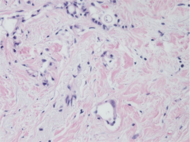
Fig. 5.

Microscopic appearance of the pancreatic tumor with hematoxylin-eosin staining. A lesion corresponding to carcinoma in situ was detected in the distal pancreatic duct, which was diagnosed as residual carcinoma invading the pancreatic duct or high-grade pancreatic intraepithelial neoplasia
