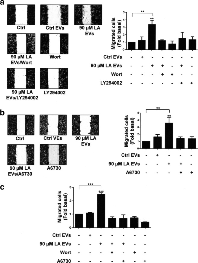
Fig. 3.

Role of PI3K and Akt in migration induced by EVs from MDA-MB-231 cells stimulated with LA in MCF10A cells. Panel a and b. Cultures of MCF10A cells were scratch-wounded and pretreated with Wort, LY294002 or A6730 and then treated with EVs from MDA-MB-231 cells untreated or treated with LA. Panel c. Migration assays were performed by using the Boyden chamber method and MCF10A cells treated with Wort and LY294002 and then stimulated with EVs from MDA-MB-231 cells untreated or treated with LA. One control of untreated MCF10A cells was included. Graphs are the mean ± S.D. and are expressed as fold of migrated cells above Ctrl EVs. Asterisks indicate comparisons made to Ctrl EVs and control. **P < 0.01, ***P < 0.001
