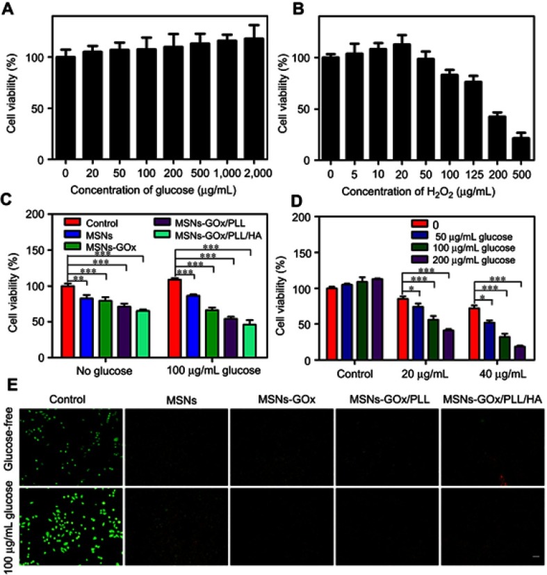
Figure 7.
Viabilities of HepG2 cells after 24 hours of incubation with different concentrations of glucose (A) and H2O2 (B). (C) The cell viability after different treatments in the absence and presence of 100 mg/mL glucose. (D) The cell viability after 24 hours of incubation with MSNs-GOx/PLL/HA (20 or 40 μg/mL) in different concentrations (0, 50, 100, 200 μg/mL) of glucose-containing DMEM media. Significant difference from control: *P<0.05, **P<0.01, ***P<0.001. (E) Fluorescent images of live and dead HepG2 cells after different treatments in glucose-free DMEM media and 100 μg/mL glucose-containing DMEM media and stained by calcein AM and PI, respectively.
Abbreviations: MSNs, mesoporous silica nanoparticles; GOx, glucose oxidase; PLL, poly (L-lysine); HA, hyaluronic acid; AM, acetoxymethyl; PI, propidium iodide.

