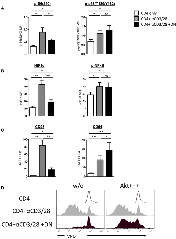
Figure 1.
DN T-cells inhibit mTOR activation but not MAPK p38 signaling in CD4 T-cells. Freshly isolated CD4 T-cells were incubated with anti-CD3/CD28 coated beads in absence (gray) or presence (black) of DN T-cells. Unstimulated CD4 T-cells were used as negative control (white). (A) Phosphorylation of ribosomal protein S6(S240) (left) and MAPK p38(T180/Y182) (right) in CD4 T-cells after 24 h culture was quantified by flow cytometry. Graphs show MFI +/-SEM of at least six independent experiments. (B) Expression of HIF-1α and NFκB(p65) was analyzed in CD4 T-cells after 24 h co-culture, graph represent MFI +/–SEM of 7 experiments. (C) Expression of CD98 and CD54 was measured after 3 days, MFI +/SEM of at least five experiments is shown. Ns, not significant, *p < 0.05, **p < 0.01, ***p < 0.01. (D) Freshly isolated VPD-labeled CD4 T-cells were incubated with SC79 (Akt+ + +) for 2 h at 37°C and washed intensively. Treated and untreated VPD-labeled CD4 T-cells were activated with anti-CD3/CD28 coated beads in presence or absence of DNT-cells for 6 days. Cells were analyzed by flow cytometry, histograms were gated for CD4 T-cells.

