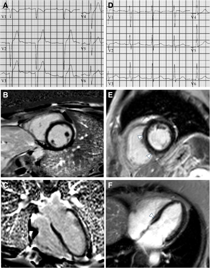
Fig. 1.
Representative ECG and CMR findings in the study cohort. Panels A shows an abnormal ECG with prolonged PR interval and borderline QRS interval of a subject with no evidence of myocardial fibrosis by CMR-LGE (B, C). Panels D shows a normal ECG of a subjects with evidence of midwall fibrosis mostly evident in the interventricular septum (arrows in E, and F)

