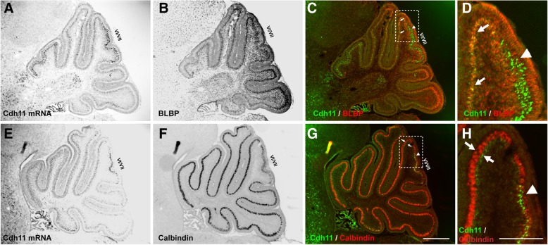
Fig. 3.
Low level of expression of Cdh11 in Burgmann glial cells. In situ hybridization of Cdh11 followed by calbindin or BLBP staining was performed on P4 sagittal sections. a-d Colocalization of BLBP signal (red in (c) and (d)) with Cdh11 in situ hybridization signal (green in (c) and (d)) in areas except the central region of lobules VI/VII is demonstrated. d The enlarged image of the boxed area in (c) shows the weak Cdh11 expression signal in BLBP-positive Burgmann glial cells. In the central area of lobules VI/VII, strong Cdh11 expression is not colocalized with BLBP signal (c and d, arrow heads). e-h a weak Cdh11 signal (green in (g) and (h)) is present underneath (inferior to) the Purkinje cell layer as indicated by calbindin signal (red in (g) and (h), arrows). Strong Cdh11 expression signal is within the calbindin-positive cell layer. Scale bar for (a-c) and (e-g): 500 μm. Scale bar for (d) and (h): 200 μm

