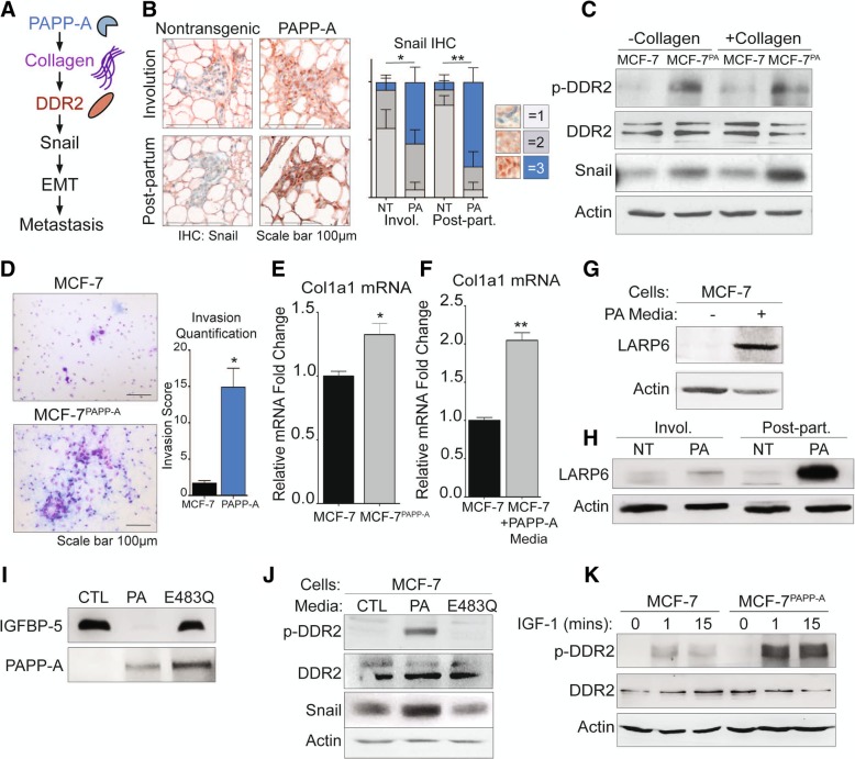
Fig. 3.
PAPP-A promotes DDR2 activation through collagen mRNA stabilization and IGF signaling in vitro. a Schematic of the proposed mechanism of activation of DDR2/Snail signaling axis of invasion [39]. b Representative images of Snail IHC (Snail: red; nuclei: blue) of virgin, involuting, or post-partum mammary glands from non-transgenic or PAPP-A transgenic mice. n = 5 mice per group. Quantification of Snail IHC is shown, n = 5 mice per group. Mean ± SEM, unpaired t test with Welch’s correction (comparisons between the score 3 group only). *p < 0.05, **p < 0.005. c Immunoblot of indicated markers in MCF-7 or MCF-7PAPP-A cells co-cultured with and without collagen. Scale bar 100 μm. d Representative images of 48 h Transwell in vitro invasion assays of MCF-7 and MCF-7PAPP-A cells. Experiments repeated in technical triplicate. Scale bar 100 μm. Quantification is shown, unpaired t test with Welch’s correction. *p < 0.05. e Relative fold change of col1a1 mRNA transcript in MCF-7PAPP-A over MCF-7 cells measured by real-time qPCR and normalized to actin. Mean + SEM, triplicate experiment. Unpaired t test with Welch’s correction. *p < 0.05. f Relative fold change of col1a1 mRNA transcript in MCF-7 cells treated in vitro with or without PAPP-A media for 24 h. PAPP-A media concentration is at 10 ng/mL of PAPP-A protein (quantified by ELISA). Measured by real-time qPCR and normalized to actin. Mean + SEM, triplicate experiment. Unpaired t test with Welch’s correction. **p < 0.005. g Immunoblot of LARP6 in MCF-7 cells treated in vitro with or without PAPP-A media for 24 h. PAPP-A media concentration is at 10 ng/mL of PAPP-A protein (quantified by ELISA). h Representative immunoblot of LARP6 in the mammary glands from non-transgenic or PAPP-A transgenic mice during virgin, involution, or late post-partum. n = 5 mice per group. i Immunoblot of rIGFBP-5 following a 3-h incubation in culture media from MCF-7 (CTL), MCF-7PAPP-A (PA), or MCF-7 overexpressing proteolytic-dead mutant PAPP-A E483Q (E483Q). Immunoblot of PAPP-A secreted in culture media from MCF-7, MCF-7PAPP-A, and MCF-7PAPP-A/E483Q cells. j Immunoblot of MCF-7 cells treated in vitro with culture media from MCF-7 (CTL), MCF-7PAPP-A (PA), or MCF-7PAPP-A/E483Q (E483Q) cells for 24 h. k Immunoblot of DDR2 and phospho-DDR2 in MCF-7 and MCF-7PAPP-A cells treated in vitro with recombinant IGF-1 at 10 nM for indicated time points

