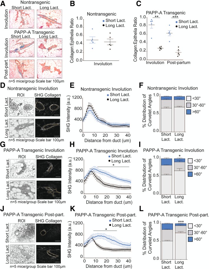
Fig. 5.
Long lactation inhibits PAPP-A-driven collagen deposition and TACS3 formation in post-partum mammary glands. a Representative images of Masson’s trichrome collagen stain (blue) on involuting or post-partum mammary glands from non-transgenic and PAPP-A transgenic mice following a short (2 days) or long (14 days) lactation period. n = 5 mice per group. All images from mice following short lactation are from the same experiment shown in Fig. 1a. Scale bar 100 μm. b Quantification of collagen per epithelial region by Masson’s trichrome stain on non-transgenic involuting mammary glands following a short (2 days) or long (14 days) lactation period from a. n = 5 mice, each point represents the average of ten ducts per mouse per time point. Mean ± SEM, unpaired t test with Welch’s correction. Analysis on short lactation samples is from the same analysis performed in Fig. 1b. c Quantification of collagen per epithelial region by Masson’s trichrome stain on PAPP-A transgenic involuting or post-partum mammary glands following a short (2 days) or long (14 days) lactation period from a. n = 5 mice, each point represents the average of ten ducts per mouse per time point. Mean ± SEM, unpaired t test with Welch’s correction: **p < 0.005, ***p < 0.0005. Analysis on short lactation samples is from the same analysis performed in Fig. 1c. d Representative second-harmonic generation (SHG) imaging of collagen on magnified ducts of histological sections of non-transgenic involuting mammary glands following a short (2 days) or long (14 days) lactation period (right panels) and corresponding regions of interest (ROI) (left panels) (n = 5 mice per group). All images of short lactation samples are from the same experiment shown in Fig. 1d. Scale bar 100 μm. e Graph of collagen intensity relative to the distance from the mammary duct borders of SHG images of non-transgenic involuting mammary glands following a short (2 days) or long (14 days) lactation period in d. Mean ± SEM. n = 5 mice per group, unpaired t test with Welch’s correction. Analysis on short lactation samples is from the same analysis performed in Fig. 1e. f Quantification of TACS3 per total curvelets analyzed in non-transgenic involuting mammary glands following a short (2 days) or long (14 days) lactation period using the CurveAlign software. TACS3 characterized as curvelet angles 60–90 relative to the ductal border. n = 5 mice per group. Mean + SEM, unpaired t test with Welch’s correction (comparisons between the TACS3 group only). Analysis on short lactation samples is from the same analysis performed in Fig. 1g. g Representative second-harmonic generation (SHG) imaging of collagen on magnified ducts of histological sections of PAPP-A transgenic involuting mammary glands following a short (2 days) or long (14 days) lactation period (right panels) and corresponding regions of interest (ROI) (left panels) (n = 5 mice per group). All images of short lactation samples are from the same experiment shown in Fig. 1d. Scale bar 100 μm. h Graph of collagen intensity relative to the distance from the mammary duct borders of SHG images of PAPP-A transgenic involuting mammary glands following a short (2 days) or long (14 days) lactation period in e. Mean ± SEM. n = 5 mice per group, unpaired t test with Welch’s correction. Analysis on short lactation samples is from the same analysis performed in Fig. 1f. i Quantification of TACS3 per total curvelets analyzed in PAPP-A transgenic involuting mammary glands following a short (2 days) or long (14 days) lactation period using the CurveAlign software. TACS3 characterized as curvelet angles 60–90 relative to the ductal border. n = 5 mice per group. Mean + SEM, unpaired t test with Welch’s correction (comparisons between the TACS3 group only). *p < 0.05. Analysis on short lactation samples is from the same analysis performed in Fig. 1g. j Representative second-harmonic generation (SHG) imaging of collagen on magnified ducts of histological sections of PAPP-A transgenic post-partum mammary glands following a short (2 days) or long (14 days) lactation period (right panels) and corresponding regions of interest (ROI) (left panels) (n = 5 mice per group). All images of short lactation samples are from the same experiment shown in Fig. 1d. k Graph of collagen intensity relative to the distance from the mammary duct edge of SHG images of PAPP-A transgenic post-partum mammary glands following a short (2 days) or long (14 days) lactation period in f. Mean ± SEM. n = 5 mice per group, unpaired t test with Welch’s correction: *p < 0.05. Analysis on short lactation samples is from the same analysis performed in Fig. 1f. l Quantification of TACS3 per total curvelets analyzed in PAPP-A transgenic post-partum mammary glands following a short (2 days) or long (14 days) lactation period using the CurveAlign software. TACS3 characterized as curvelet angles 60–90 relative to the ductal border. n = 5 mice per group. Mean + SEM, unpaired t test with Welch’s correction (comparisons between the TACS3 group only). *p < 0.05. Analysis on short lactation samples is from the same analysis performed in Fig. 1g

