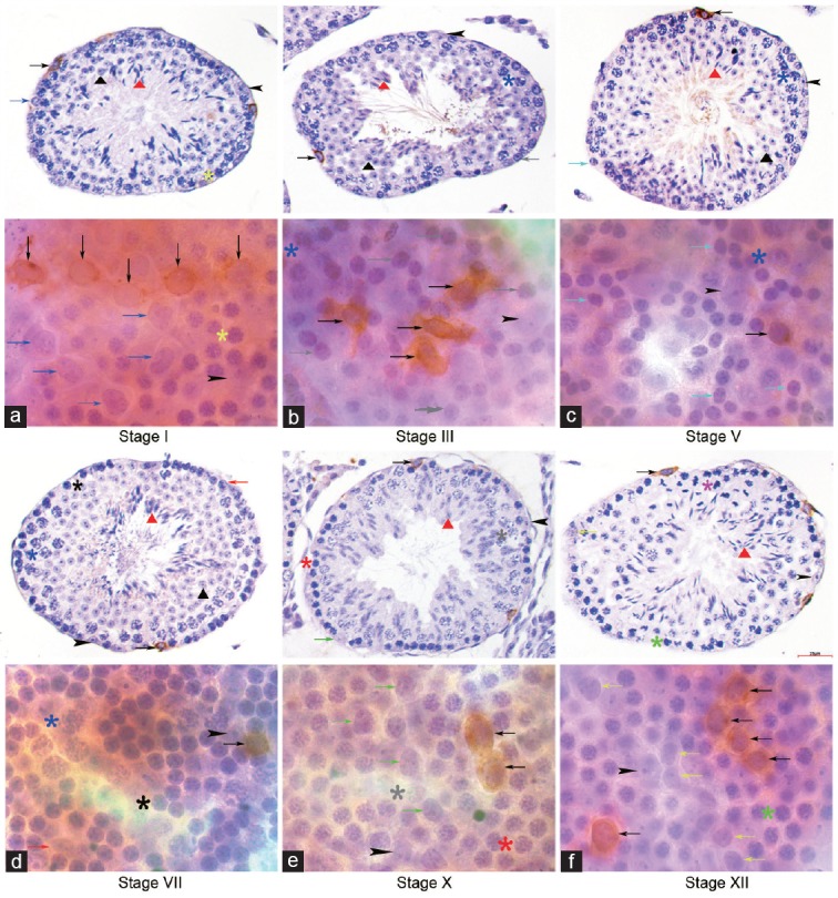
Figure 3.

Expression profile of GFRA1 in cross section and whole mount IHC. GFRA1 immuno-staining was detected mostly on the cytomembrane of As, Apr and Aal4 undifferentiated spermatogonia clones across the seminiferous cycle. No GFRA1 staining was detected in (a) A4 in stage I, (b) In in stage III, (c) B in stage V, (d) A1 in stage VII, (e) A2 in stage X and (f) A3 in stage XII in cross section and whole mount IHC. Indicators of multiple germ cell types were the same as in figure 1. Scale bar=25 μm.
