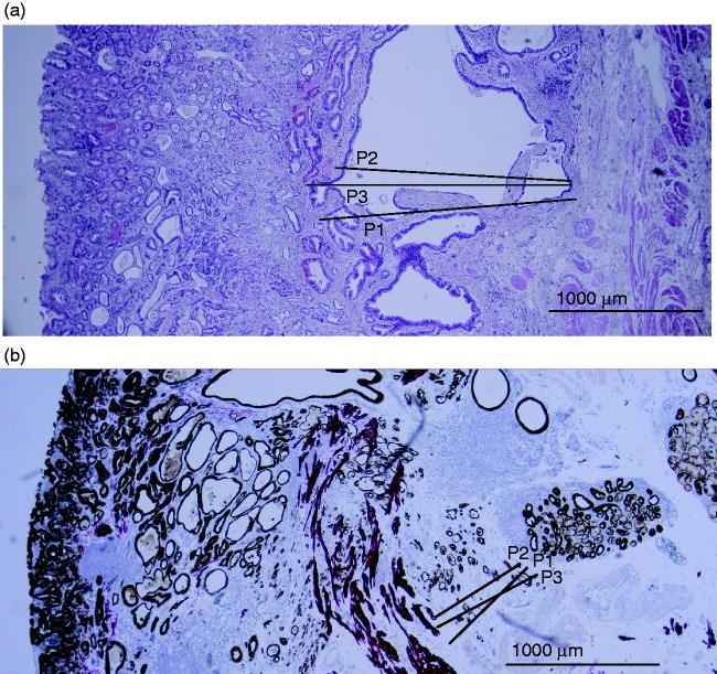
Figure 1.
Histological assessment of pT1b OAC, scored independently by three gastrointestinal pathologists. (a) Surgical resection specimen, stained by hematoxylin and eosin: all three pathologists scored a moderate grade of differentiation (G2) and no lymphovascular invasion (LVI). Submucosal invasion depth for pathologist 1 (P1) was 1720 µm, pathologist 2 (P2) was 1525 µm and pathologist 3 (P3) was 1890 µm. (b) Endoscopic resection specimen, with desmin and pankeratin immunohistochemistry: all three pathologists scored no LVI. The grade of differentiation was moderate for P1, good for P2 and moderate for P3. Submucosal invasion depth for P1 was 581 µm, P2 was 340 µm and P3 was 620 µm.

