ABSTRACT
Background
Digital revolution is here and becoming more and more influential in our daily lives by transforming several things such as our habits, interactions with other people and the practice of dentistry. In implant dentistry, the newer methods by using cone-beam computed tomography and computer-aided design and computer-aided manufacturing recently offer more predictable and aesthetics outcomes in a shorter period of treatment time when compared to the traditional prosthetic procedures.
Methods
A 66 year-old male patient with an edentulous mandible and several failing maxillary teeth presented to our clinic. After cone-beam computed-tomography scans and virtual implant placement by using three-dimensional software, a stereolithographic surgical guide was fabricated. The patient received two mandibular implants without any flap elevation by means of a computer-aided design and computer-aided manufacturing surgical guide and a maxillary complete denture in a day.
Results
The surgical and restorative procedures were performed without any issues. The patient was followed-up for three years and no major complications with the implants and prostheses were observed.
Conclusions
The technique illustrated in this report may be successfully used to restore edentulous arches in a day if it is executed by trained restorative dentists and if patient selection is appropriate.
Keywords: cone-beam CT, CAD/CAM, implant, overdenture, stereolithography, surgery
INTRODUCTION
Each year, new technologies emerge in the dental world that change and improve existing workflows [1-4]. While some innovations are geared toward improved and faster usability, some new solutions are so big that the promise for drastic change is felt immediately. One of the remarkable things about dental technology is how much it borrows from other industries-like computer-aided design and computer-aided manufacturing (CAD/CAM) which came from the manufacturing sector or cone-beam computed tomography (CBCT) which was pioneered in the late 1990s [5-7]. Both of these technologies changed implantology field drastically through many advantages they offer to both clinicians and patients.
CAD/CAM using stereolithography method has had a huge impact in implant dentistry by enabling fabrication of implant surgical guide to allow for accurate transfer of planned implant sites to the actual patient’s mouth [5,6]. Therefore, the precision of implant surgical phase increases and chances of surgical error diminishes. It can be concluded that technologies that were invented without a thought to dentistry have been pulled over and changed dentistry particularly implant dentistry forever.
In the dental literature, a few clinical reports presenting successful results with CAD/CAM surgical guides and immediate loading are available [8-11], but there is no clinical report that flapless surgery, CAD/CAM surgical guide, duplication procedure, and implant-retained mandibular overdenture were used in the same patient in a day. The objectives of this report were to describe the technique, in which an edentulous mandible was rehabilitated in a day by using an immediately-loaded implant-retained overdenture with flapless surgery and a CAD/CAM surgical guide and to present the 3-year outcomes.
CASE DESCRIPTION AND RESULTS
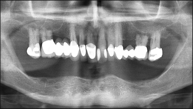
A 66-year old male patient with a mandibular complete denture and several maxillary teeth with periodontal problems presented to our faculty practice. His chief complaint was poor aesthetics and diminished masticatory efficacy and denture retention. A series of meticulous clinical and radiographic evaluations including CBCT scans were carried out (Figure 1). The patient could not afford an implant-supported fixed prosthesis, but accepted prosthodontically-driven treatment plan: the placement of two implants in the anterior mandible without any soft tissue flap elevation, an immediately-loaded implant-retained mandibular overdenture immediately, and the extraction of all existing maxillary teeth and a maxillary complete denture. The following steps were adhered to restore edentulous mandible and hopeless maxillary teeth.
Figure 1.

Presurgical panoramic radiograph of the patient.
The presurgical steps:
Eight fiducial markers (gutta-percha) were inserted into the interim mandibular denture to use it as a radiographic guide. Two sets of CBCT (3D Accuitomo, Morita, Kyoto, Japan) scan were obtained; a) while the patient was having the guide intraorally and stabilizing with an occlusal registration record, b) while the guide was on the scanning table after removal from the mouth. The settings for CBCT machine were 90 kV, 5 mA, 18 s, 360° rotation, 250 μm isotropic voxel size, interval distance 250 μm, field of view 8 x 8 cm.
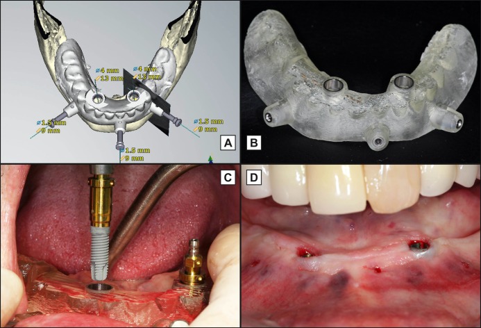
Two sets of images were saved in digital imaging and communications in medicine (DICOM) format on a computer. These DICOM images were transferred into the three-dimensional implant planning software NobelClinician software (Nobel Biocare, Yorba Linda, CA, USA). Two mandibular implants were virtually placed with respect to the treatment plan. (Figure 2A). Once completed, the data of the virtual plan were electronically transmitted to the production centre in standard tessellation language format.
After the fabrication of the mandibular surgical guide (Figure 2B), its fit was intraorally verified. The occlusal record was modified as needed with a fast-setting polyvinyl siloxane material.
Figure 2.
A = occlusal view of the completed virtual implant placement and computer-aided design and computer-aided manufacturing (CAD/CAM) surgical guide.
B = CAD/CAM surgical guide after fabrication.
C = flapless implant placement through the metal sleeves of the CAD/CAM surgical guide.
D = intraoral view of two mandibular implants immediately after implant placement.
The day of surgery:
Local anaesthesia was achieved after getting the patient’s informed consent. The mandibular surgical guide was inserted in the mouth and secure it by means of three stabilization pins. The implants (4 x 13 mm, NobelReplace Straight Groovy, Nobel Biocare, Yorba Linda, CA, USA) were placed through the metal sleeves in the surgical guide (NobelGuide, Nobel Biocare, Yorba Linda, CA, USA) with a flapless surgical approach (Figure 2C, D) after completing the implant socket preparations.
Immediately after the implant placement, the primary implant stability was measured by using resonance frequency analysis (Osstell, Integration Diagnostics, Göteborg, Sweden). Implant stability quotient values over 65, which is a prerequisite for the immediate loading should be accomplished [12]. ISQ values were 87 and 89 for the left and right implants in this case respectively.
Two locator abutments (Zest Anchors, Escondido, CA, USA) were inserted on the implants and the metal housings (Zest Anchors, Escondido, CA, USA) were seated on the locators. The existing mandibular denture was relieved to generate room for both locators and metal housings.
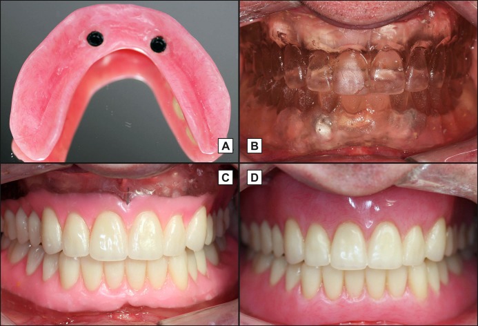
The patient’s existing mandibular complete denture was converted to an immediately loaded implant-retained overdenture by attaching the metal housings to the denture with autopolymerizing acrylic resin (Lang Dental, Wheeling, IL, USA) (Figure 3A).
All existing hopeless maxillary teeth were extracted and extraction sockets were sutured. The previously fabricated maxillary interim denture was inserted. Tissue conditioning may be needed at this moment. The patient was dismissed for the day after giving the postsurgical instructions.
Figure 3.
A = internal view of the implant-retained mandibular overdenture.
B = intraoral view of the patient after both duplicate trial dentures were inserted.
C = intraoral view of the patient with new denture teeth were arranged.
D = intraoral view of the patient after both maxillary complete denture and implant-retained mandibular overdenture were delivered.
A month later:
The interim maxillary complete denture and implant-retained mandibular overdenture were duplicated by using condensation silicone putty (Sil-Tech, Ivoclar, Amherst, NY, USA) and autopolymerizing acrylic resin (Lang Dental, Wheeling, IL, USA).
Both duplicate dentures were inserted intraorally and occlusal vertical dimension and centric relation (Figure 3B) were confirmed. Any undercuts were blocked out in the intaglio surface of the duplicate dentures. Stone was poured under the dentures to create definitive casts after applying a separating medium to the intaglio surface.
Both casts with duplicate dentures were mounted on a semi-adjustable articulator (Whip-mix, Louisville, KY, USA). The clear teeth on the duplicate dentures were eliminated by grinding, and new denture teeth (Ivoclar Vivadent, Amherst, NY, USA) were arranged on the duplicate dentures (Figure 3C).
The maxillary and mandibular duplicate dentures with new teeth were inserted in the mouth, and occlusal vertical dimension, phonetics and aesthetics were corroborated. Sufficient clearance for the locators and metal housings were accomplished under the overdenture. If needed, a final impression can be made by relining the duplicate dentures by using a polyvinyl siloxane impression material and closed-mouth approach at this moment.
Both dentures were processed, finished, and polished in the laboratory. The two metal housings were attached to the mandibular denture with autopolymerizing acrylic resin and an implant-retained mandibular overdenture was fabricated (Figure 3D). Lastly, both restorations were intraorally inserted and any necessary adjustments were made.
After the implant placement, the patient did not experience any swelling or soft tissue complications and had minimal discomfort that was managed by pain relievers in a day. A rapid healing was observed as no soft tissue flap was raised. During the follow-up period of 3 years, both implants were stable and showed peri-implant bone loss of about 1.5 mm. The maxillary complete denture was relined in the laboratory 6 month after the surgery, and the plastic retentive inserts were replaced in the clinic about a year later but no other adjustments/repairs were made.
DISCUSSION
CAD/CAM technology has become popular in surgical and restorative parts of implant dentistry [13-15]. A full-thickness gingival flap is traditionally elevated to see the surgical sites for dental implant placement. In addition, flap elevation allows that certain anatomical structures such as maxillary sinuses and mental foramina, are easily identified and protected. However, flap elevation requires suturing, and is associated with some gingival recession, bone resorption, morbidity and discomfort [16]. The technique of flapless implant surgery has been suggested for the patients to minimize the possibility of postoperative peri-implant tissue loss and to overcome the challenge of soft tissue management after surgery [16-18]. A few previous clinical reports indicated that the flapless implant placement resulted in less pain with shorter duration compared to patients in whom conventional flaps were raised [18].
Although an edentulous mandible was successfully restored in a day by using a flapless surgical method via a CAD/CAM surgical guide in the present report, there are many reports presenting certain level of deviation [5-19]. In a clinical study by Ozan et al. [19], 110 implants were inserted by utilizing CAD/CAM surgical guides that used the data from computed tomography. They observed that the mean angular deviation of all placed implants was 4.1 degrees, and the mean linear deviation was 1.11 mm at the implant platform and 1.41 mm at the implant apex when compared to the planned implants. It is essential to mention that there is a learning curve for this method. It is recommended that novice clinicians who want to use this technique should obtain advanced training [20]. Otherwise, this method may not yield predictable outcomes.
A clinical study by Valente et al. [21] searched the accuracy of guided-implant surgery by comparing the positions of planned and placed implants. Their study included 104 implants in 25 patients, which were placed by using computerized tomography, and CAD/CAM surgical guides. Only 89 implants were used to make accuracy comparisons in their study. They reported that mean lateral deviations at the coronal and apical ends of the implants were 1.4 mm and 1.6 mm. They also observed that the mean depth and angular deviations were 1.1 mm and 7.9 degrees. In another clinical study, Vieira et al. [22], assessed the accuracy of a flapless implant placement by using CAD/CAM surgical guides. Sixty two implants were placed in 14 patients with the help of stereolithographic surgical guides. They reported that the actual placed implants, when compared to the planned implants, demonstrated mean linear deviations at the cervical, middle, and apical implant sections of 2.17 mm, 2.32 mm, and 2.86 mm for the maxilla; and 1.42 mm, 1.42 mm, and 1.42 mm for the mandible, respectively.
CONCLUSIONS
This report portrays how to restore an edentulous mandible with an implant-retained mandibular overdenture by using a flapless surgical method by means of a computer-aided design and computer-aided manufacturing surgical guide in a day. This treatment modality may present predictable outcomes especially in the anterior mandible where accomplishing high primary implant stability is easier.
Acknowledgments
ACKNOWLEDGMENTS AND DISCLOSURE STATEMENTS
There are no conflicts of interest related to this study.
The author thanks Dr. William F. Skiba for his help in proofreading this article.
REFERENCES
- 1.Van Assche N, Fickl S, Francisco H, Gurzawska K, Milinkovic I, Navarro JM, Torsello F, Thoma DS. Guidelines for development of Implant Dentistry in the next 10 years regarding innovation, education, certification, and associations. Clin Oral Implants Res. 2018 Jun;29(6):568-575. [DOI] [PubMed]
- 2.Turkyilmaz I, Asar NV. Eighteen-Month Outcomes of Titanium Frameworks Using Computer-Aided Design and Computer-Aided Manufacturing Method. Implant Dent. 2017 Jun;26(3):480-484. [DOI] [PubMed]
- 3.Dano D, Stiteler M, Giordano R. Prosthetically Driven Computer-Guided Implant Placement and Restoration Using CEREC: A Case Report. Compend Contin Educ Dent. 2018 May;39(5):311-317. [PubMed]
- 4.Jorba-García A, Figueiredo R, González-Barnadas A, Camps-Font O, Valmaseda-Castellón E. Accuracy and the role of experience in dynamic computer guided dental implant surgery: An in-vitro study. Med Oral Patol Oral Cir Bucal. 2019 Jan 1;24(1):e76-e83. [DOI] [PMC free article] [PubMed]
- 5.Turbush SK, Turkyilmaz I. Accuracy of three different types of stereolithographic surgical guide in implant placement: an in vitro study. J Prosthet Dent. 2012 Sep;108(3):181-8. [DOI] [PubMed]
- 6.Vasak C, Kohal RJ, Lettner S, Rohner D, Zechner W. Clinical and radiological evaluation of a template-guided (NobelGuide™) treatment concept. Clin Oral Implants Res. 2014 Jan;25(1):116-23. [DOI] [PubMed]
- 7.Neumeister A, Schulz L, Glodecki C. Investigations on the accuracy of 3D-printed drill guides for dental implantology. Int J Comput Dent. 2017;20(1):35-51. [PubMed]
- 8.Abboud M, Wahl G, Calvo-Guirado JL, Orentlicher G. Application and success of two stereolithographic surgical guide systems for implant placement with immediate loading. Int J Oral Maxillofac Implants. 2012 May-Jun;27(3):634-43. [PubMed]
- 9.Shen P, Zhao J, Fan L, Qiu H, Xu W, Wang Y, Zhang S, Kim YJ. Accuracy evaluation of computer-designed surgical guide template in oral implantology. J Craniomaxillofac Surg. 2015 Dec;43(10):2189-94. [DOI] [PubMed]
- 10.Nikzad S, Azari A. Custom-made radiographic template, computed tomography, and computer-assisted flapless surgery for treatment planning in partial edentulous patients: a prospective 12-month study. J Oral Maxillofac Surg. 2010 Jun;68(6):1353-9. [DOI] [PubMed]
- 11.Scotti R, Pellegrino G, Marchetti C, Corinaldesi G, Ciocca L. Diagnostic value of NobelGuide to minimize the need for reconstructive surgery of jaws before implant placement: a review. Quintessence Int. 2010 Nov-Dec;41(10):809-14. [PubMed]
- 12.Turkyilmaz I, McGlumphy EA. Is there a lower threshold value of bone density for early loading protocols of dental implants? J Oral Rehabil. 2008 Oct;35(10):775-81. [DOI] [PubMed]
- 13.Tallarico M, Meloni SM, Canullo L, Caneva M, Polizzi G. Five-Year Results of a Randomized Controlled Trial Comparing Patients Rehabilitated with Immediately Loaded Maxillary Cross-Arch Fixed Dental Prosthesis Supported by Four or Six Implants Placed Using Guided Surgery. Clin Implant Dent Relat Res. 2016 Oct;18(5):965-972. [DOI] [PubMed]
- 14.Testori T, Robiony M, Parenti A, Luongo G, Rosenfeld AL, Ganz SD, Mandelaris GA, Del Fabbro M. Evaluation of accuracy and precision of a new guided surgery system: a multicenter clinical study. Int J Periodontics Restorative Dent. 2014;34 Suppl 3:s59-69. [DOI] [PubMed]
- 15.Tahmaseb A, De Clerck R, Aartman I, Wismeijer D. Digital protocol for reference-based guided surgery and immediate loading: a prospective clinical study. Int J Oral Maxillofac Implants. 2012 Sep-Oct;27(5):1258-70. [PubMed]
- 16.Campelo LD, Camara JR. Flapless implant surgery: a 10-year clinical retrospective analysis. Int J Oral Maxillofac Implants. 2002 Mar-Apr;17(2):271-6. [PubMed]
- 17.Rocci A, Martignoni M, Gottlow J. Immediate loading in the maxilla using flapless surgery, implants placed in predetermined positions, and prefabricated provisional restorations: a retrospective 3-year clinical study. Clin Implant Dent Relat Res. 2003;5 Suppl 1:29-36. [DOI] [PubMed]
- 18.Fortin T, Bosson JL, Isidori M, Blanchet E. Effect of flapless surgery on pain experienced in implant placement using an image-guided system. Int J Oral Maxillofac Implants. 2006 Mar-Apr;21(2):298-304. [PubMed]
- 19.Ozan O, Turkyilmaz I, Ersoy AE, McGlumphy EA, Rosenstiel SF. Clinical accuracy of 3 different types of computed tomography-derived stereolithographic surgical guides in implant placement. J Oral Maxillofac Surg. 2009 Feb;67(2): 394-401. [DOI] [PubMed]
- 20.Fernández-Gil Á, Gil HS, Velasco MG, Moreno Vázquez JC. An In Vitro Model to Evaluate the Accuracy of Guided Implant Placement Based on the Surgeon's Experience. Int J Oral Maxillofac Implants. 2017 May/Jun;32(3):151-154. [DOI] [PubMed]
- 21.Valente F, Schiroli G, Sbrenna A. Accuracy of computer-aided oral implant surgery: a clinical and radiographic study. Int J Oral Maxillofac Implants. 2009 Mar-Apr;24(2):234-42. [PubMed]
- 22.Vieira DM, Sotto-Maior BS, Barros CA, Reis ES, Francischone CE. Clinical accuracy of flapless computer-guided surgery for implant placement in edentulous arches. Int J Oral Maxillofac Implants. 2013 Sep-Oct;28(5):1347-51. [DOI] [PubMed]


