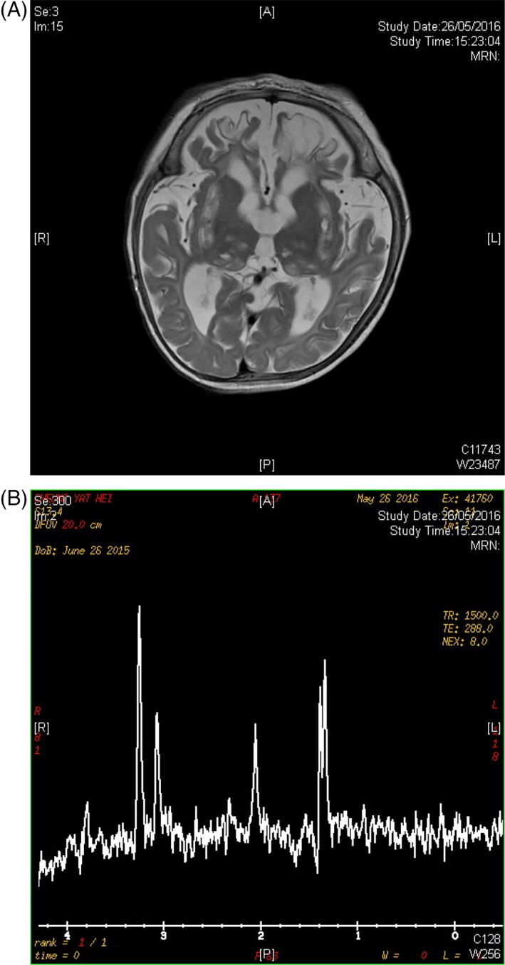
Figure 1.

Magnetic resonance imaging brain images of the index subject. A, T2W hyperintense cystic changes involving bilateral corona radiata, basal ganglia and thalami, compatible with old lacunar infarcts, cerebral atrophy with encephalomalacic changes in bilateral frontal lobes. B, Doublet lactate peaks on magnetic resonance spectroscopy
Abbreviation: MRI, magnetic resonance imaging
