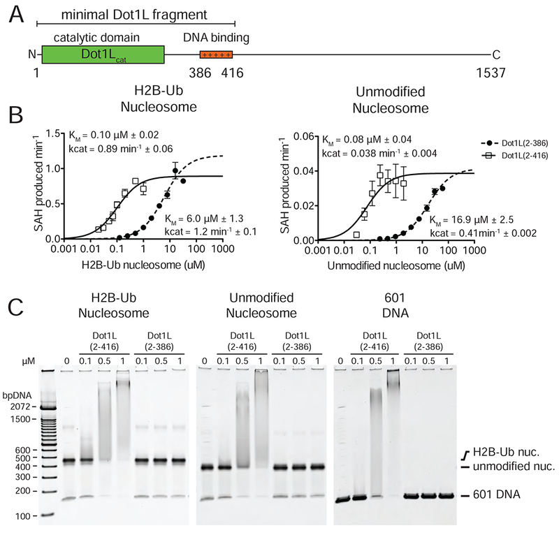
Figure 2 : Analysis of Dot1L binding and activity.

A, Schematic of the Dot1L domain architecture. B Michaelis Menten titrations of two variants of Dot1L. Left, titration with H2B-Ub nucleosome. Right, titration with unmodified Nucleosome. Error bars correspond to the standard error of three replicate measurements. The kcat and Km of the fitted data are reported in the graph field. The reported errors of the fitted Km and kcat correspond to the standard error. C Electrophoretic Mobility Shift Assay of Dot1L variants binding to different nucleosome substrates and to free DNA.
