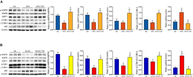
FIGURE 3.

TSF enhanced autophagy and increased AMPK activation and SIRT1 expression in mice fed a HFD or MCDD. (A) Western blot assay and semi-quantitative analysis of p-AMPK, AMPK, SIRT1, LC3B-II, p62 expression and the ratio of LC3B-II to LC3B-I in mice fed a HFD treated with or without TSF; data from each group are expressed as the mean ± SEM (n = 6) from three repeated western blot experiments of each mouse. (B) Western blot assay and semi-quantitative analysis of p-AMPK, AMPK, SIRT1, LC3B-II, and p62 expression in mice fed a MCDD treated with or without TSF; data from each group are expressed as the mean ± SEM (n = 6) from three repeated western blot experiments of each mouse. aP < 0.05, bP < 0.01 vs. ND group; cP < 0.05, dP < 0.01 vs. MCDD group.
