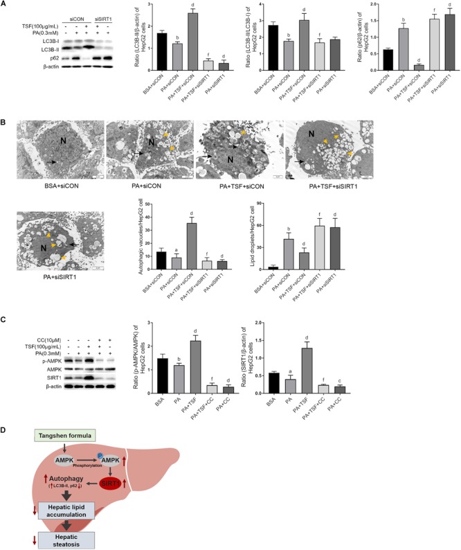
FIGURE 7.

TSF-induced autophagy is AMPK/SIRT1 dependent in PA-stimulated HepG2 cells. (A) Knockdown SIRT1 by siRNA abolished TSF-induced upregulation of LC3B-II, the ratio of LC3B-II to LC3B-I and downregulation of p62 in PA-stimulated HepG2 cells, as determined by western blot assay and semi-quantitative analysis, data are expressed as the mean ± SEM of three independent experiments performed in duplicate. (B) Knockdown SIRT1 by siRNA abolished TSF-induced upregulation of autophagic vacuoles with double-membrane structure (black arrows) and reduction in lipid droplets (yellow arrows) in PA-stimulated HepG2 cells, as determined by transmission electron microscopy; N represents the nucleus; bar = 2 μm; the number of autophagic vacuoles and lipid droplets per cell (n = 30) was counted. (C) Inhibiting AMPK phosphorylation via compound C abolished TSF-induced upregulation of SIRT1, as determined by western blot assay and semi-quantitative analysis, data are expressed as the mean ± SEM of three independent experiments performed in duplicate. (D) Model of the newly proposed mechanism in whereby TSF signaling alleviates hepatic steatosis by inducing AMPK/SIRT1 pathway mediated autophagy.aP < 0.05, bP < 0.01 vs. BSA group; dP < 0.01 vs. PA group; fP < 0.01 vs. PA+TSF group.
