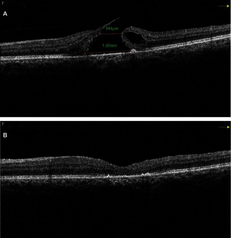
Figure 3.
(A) Pre-operative SD OCT of a large MH with a horizontal linear width of 544 μm and base diameter of 1,200 μm. (B) Post-operative SD OCT of the same patient shows a U-type successful closure after vitrectomy with the inverted ILM flap technique.
Abbreviations: SD OCT, sSpectral domain optical coherence tomography; MH, mMacular hole.

