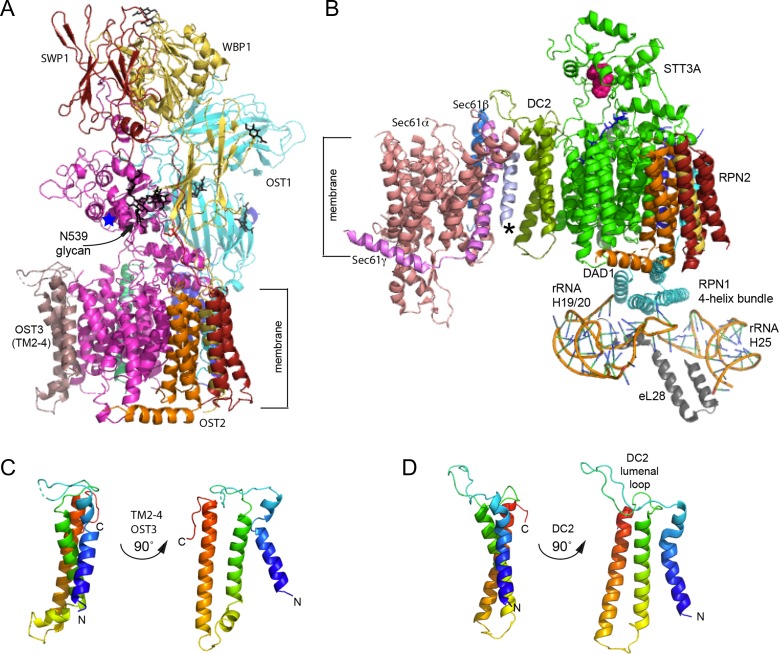
Fig. 2.
Structure of the eukaryotic oligosaccharyltransferases. (A) High-resolution structure of the yeast OST viewed from the plane of the membrane with the ER lumenal domain at the top. Subunits are color coded as in Figure 1 and are labeled. The blue star is positioned near the WWD motif. N-glycans are shown as black sticks. (B) Interaction between the STT3A complex and the RNC-Sec61 complex shown in the same orientation as in panel A. OST subunits are color coded as in Figure 1. The lumenal loop of DC2 interacts with the C-terminal tails of Sec61β (marine) and Sec61γ (light magenta). Lumenal domains of RPN1, RPN2, OST48 and TMEM258 are not shown. The C-terminal four-helix bundle of RPN1 interacts with 60 S ribosomal subunit protein eL28 and 28 S rRNA helices H25 and H19/20. The WWD motif in STT3A is shown as magenta spheres. An unidentified TM span near Sec61 is designated by an asterisk. (C, D) Structural homology between TMs 2-4 of OST3 (C) and DC2 (D) as viewed from the plane of the membrane. N and C-termini are labeled, where N of OST3 corresponds to residue S216. The figure was made with PYMOL v2.1software and PDB files 6FTI (B) and 6EZN (A, C).

