Figure 1.
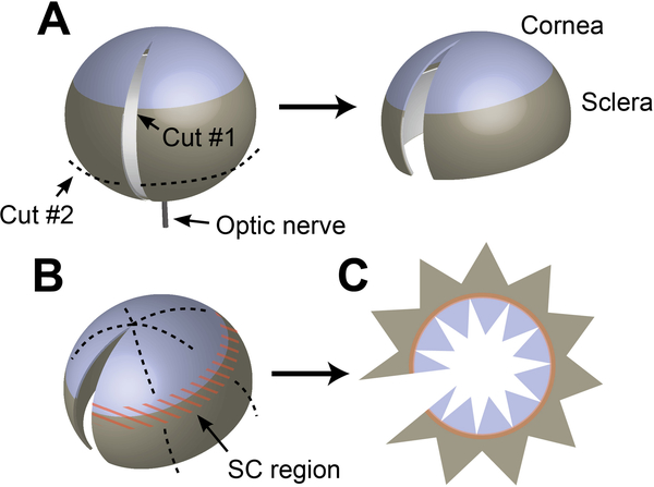
Preparing enucleated eyes for staining and mounting. (A) After removing the conjunctiva and other connective tissue from the surface of the globe, remove the optic nerve by cutting flush with the surface of the eye with scissors. While grasping the eye through the hole left by the optic nerve, a razor blade is used to make an incision from the optic nerve to the center of the cornea. A second cut (dashed line) is then made to remove the lower third of the sclera. (B) After staining, scissors are used to make a series of cuts in the sclera and cornea in preparation for flat mounting—taking care to avoid cutting into the iridocorneal angle/limbal region (orange). (C) After cutting, the eye is opened and laid flat on a glass slide in preparation for mounting.

