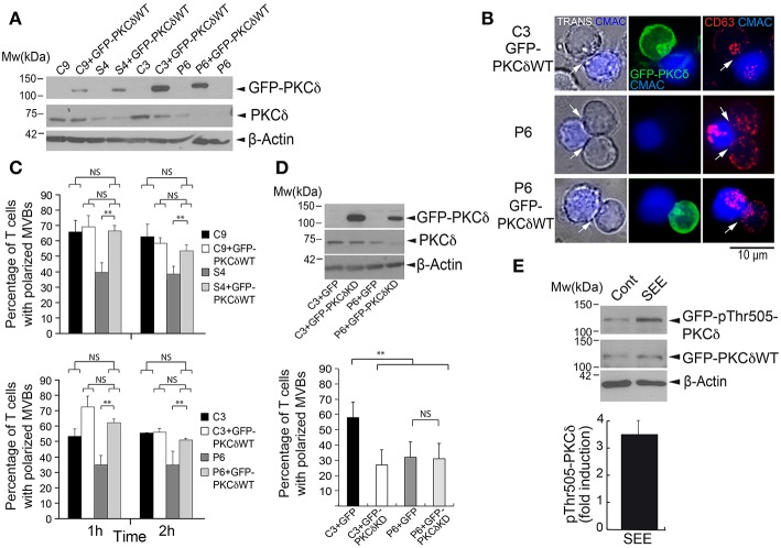
Figure 2.
GFP-PKCδ expression restores the polarization of MVB. C3 and C9 control and S4 and P6 PKCδ-interfered Jurkat clones were transfected or not with GFP, GFP-PKCδWT, or GFP-PKCδKD mutant. (A) WB of lysates from C3, C9, S4, and P6 clones, transfected or not with GFP-PKCδWT, was carried out with an anti-ratPKCδ, anti-humanPCKδ and anti-β-actin Abs. (B) Cells were challenged with CMAC-labeled SEE-pulsed Raji cells to induce synaptic conjugate formation. After 1 h cells were fixed, permeabilized, stained with anti-CD63 Abs and imaged by fluorescence microscopy. Transmittance (TRANS) plus CMAC is shown in the left panel, and the white arrows indicate the IS areas. CMAC labeling of Raji cells in blue, GFP-PKCδ in green and CD63 in red. Representative examples of polarized C3 GFP-PKCδWT+ clone, non-polarized P6 clone and polarized P6 GFP-PKCδWT+ clone are shown. Scale bar, 10 μm. (C) Quantification of MVB polarization in C9, C3 (control) and S4, P6 (PKCδ-interfered) Jurkat clones transfected or not with GFP-PKCδWT and challenged with CMAC-labeled, SEE-pulsed Raji cells for 1 and 2 h. The percentage of synapse-forming clones with polarized MVB, expressing or not expressing GFP-PKCδWT, was determined as indicated in Materials and Methods section (see also Supplementary Figure 1). Data are means plus SD (n = 3, analyzing at least 50 synapses from 15 different microscopy fields per experiment). Single-factor ANOVA was performed between the indicated groups. NS, not significant, **p ≤ 0.05. (D) Upper panel: Lysates of C3 and P6 clones transfected with either GFP or GFP-PKCδKD mutant were analyzed by WB with an anti-ratPKCδ, anti-humanPKCδ and anti-β-actin Abs. Lower panel: quantification of MVB polarization efficiency in C3 control and P6 PKCδ-interfered Jurkat clones transfected with either GFP or GFP-PKCδKD and challenged with CMAC-labeled, SEE-pulsed Raji cells for 1 h. The percentage of synapse-forming clones with polarized MVB, expressing or not expressing GFP-PKCδKD, was determined as in (C) Data are means plus SD (n = 3, analyzing at least 40 synapses from 15 different microscopy fields per experiment) and single-factor ANOVA was performed between the indicated groups. NS, not significant, **p ≤ 0.05. (E) C3 control Jurkat clone was transfected with GFP-PKCδWT and challenged with non-pulsed (Cont) or SEE-pulsed (SEE) Raji cells for 1 h. Cells were lysed and lysates analyzed by WB with anti-phosphoThr505-PKCδ, anti-rat PKCδ Ab (C-17) and anti-β-actin to normalize. In the lower panel, mean fold induction plus SD (n = 3) of normalized phosphoThr505-PKCδ signal is represented.

