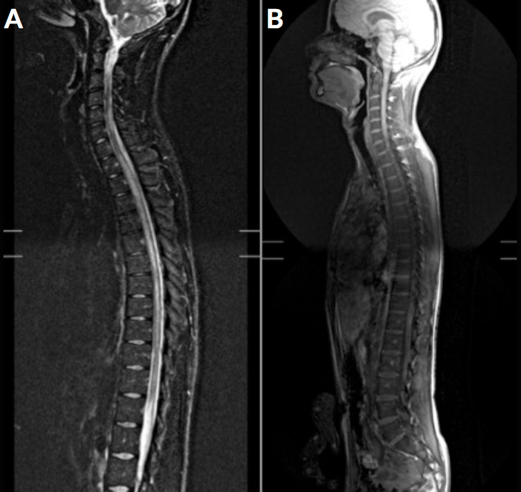
Figure 1.

A. T2R-weighted medullary MRI in the sagittal plane: medullary lesions of centromedullary topography (white arrows) and hyperintensity extending over the entire cervical-dorsal marrow to the cone terminal. Normal calibre of the spinal cord.
B. After injection of contrast product, on the appearance of some medullary lesions. No sign of epiduritis.
