
Figure 2.
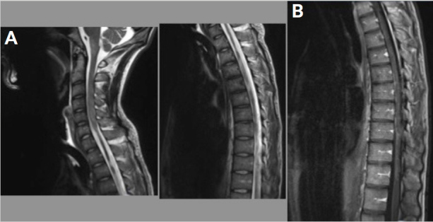
A. T2-weighted TSE-medial MRI shows intramedullary lesions in diffuse hyperintensities with atrophy of the cervical-dorsal cord. B. After intravenous injection of contrast medium, diffuse meningeal enhancement, no enhancement of intramedullary lesions.