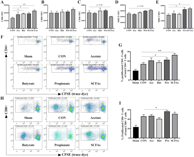
FIGURE 5.
Effects of SCFAs on the expression of surface markers (A) CD86, (B) CD80, (C) CD40, (D) MHC-II, and (E) MHC-I, and percentage of proliferated (G) CD4+ and (I) CD8+ T cells after ex vivo restimulation. Maturation status of different treated BMDCs was distinguished based on their expression (MFI) of surface markers. Representative plots of proliferated (F) CD4+ and (H) CD8+ T cell after ex vivo restimulation by different BMDCs. The Kruskal-Wallis nonparametric test, followed by Dunn's post-hoc test for selected pairs, was used for panels A–E, G, and I. Data are presented as mean ± SEM, n = 4 for panels A–E; for panels G and I, spleens were obtained from 3 nonvaccinated (sham) or 6 vaccinated mice; BMDCs were obtained from 3 donor mice. Significantly different from CON: *P < 0.05; **P < 0.01. Ace, acetate; BMDC, bone marrow–derived dendritic cell; But, butyrate; CON, control; MFI, medium fluorescence intensity; Pro, propionate.

