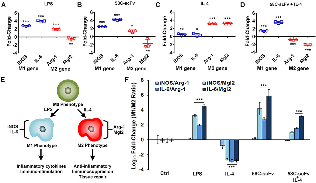
Figure 5. Effect of free, monomeric 58C-scFv on macrophage polarization.
iNOS and IL-6 were used as M1 markers and Arg-1 an Mgl2 were used as M2 markers. Raw 264.7 macrophages treated with (A) LPS, (B), 58C-scFv, (C), IL-4, and (D) 58C-scFv + Il4. In A-C the genes values were normalized to the untreated control, where D was normalized to the IL-4 only control. (E) Schematics of macrophage polarization. (F) Ratios of M1/M2 marker genes across different conditions normalized to untreated control. Data are represented as mean ± sd, *p<0.05, **p<0.01, ***p<0.001 with one-way ANOVA followed by Dunnett’s post-test.

