
Figure 5.
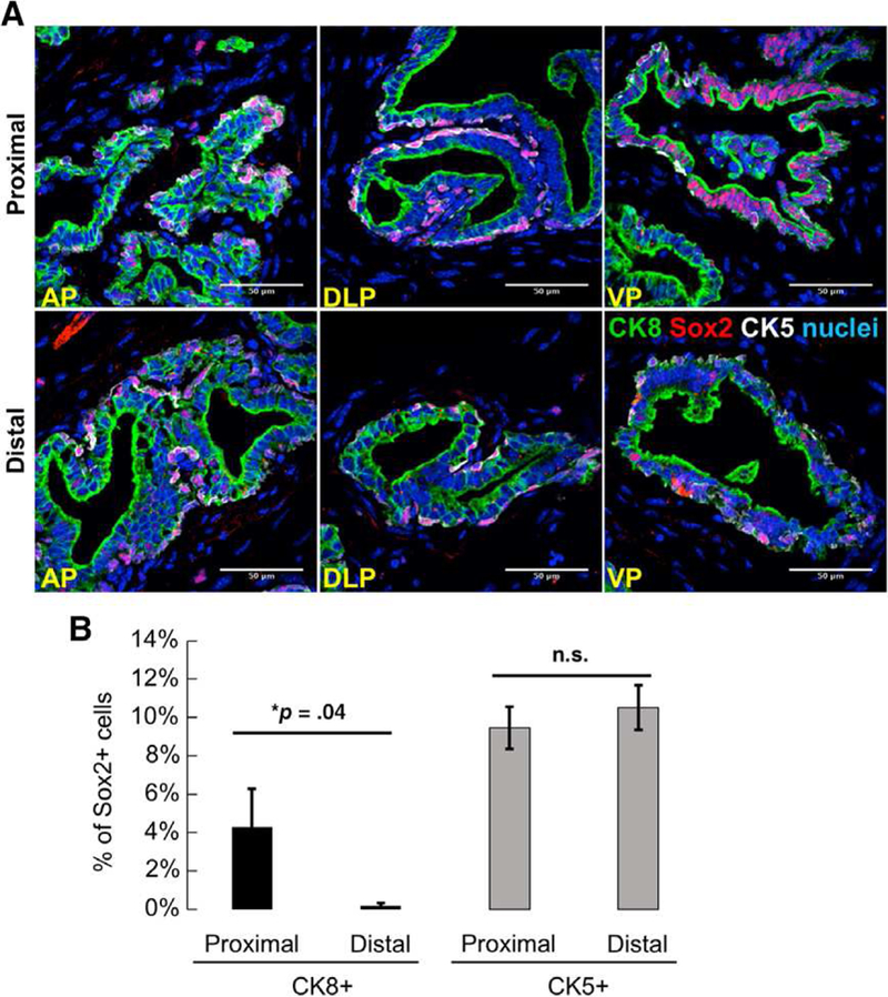
Persisting luminal cells express Sox2 after host castration. (A): Immunofluorescent staining shows colocalization of Sox2 with CK5 (white) or CK8 (green) in postpubescent castrated murine prostates (n = 3 mice, 3-weeks postcastration). Yellow arrows: Sox2+ cells positive for basal cell markers. White arrows: Sox2+ cells positive for luminal cell markers. Scale bar: 50 μm. Abbreviations: AP, anterior prostate; DLP, dorsolateral prostate, VP, ventral prostate. (B): Quantitation of Sox2+ cells in proximal and distal regions of prostates from adult, castrated mice. Data represent the mean T SEM, one-tailed homoscedastic Student’s t test; *, p = .04.