
Figure 6.
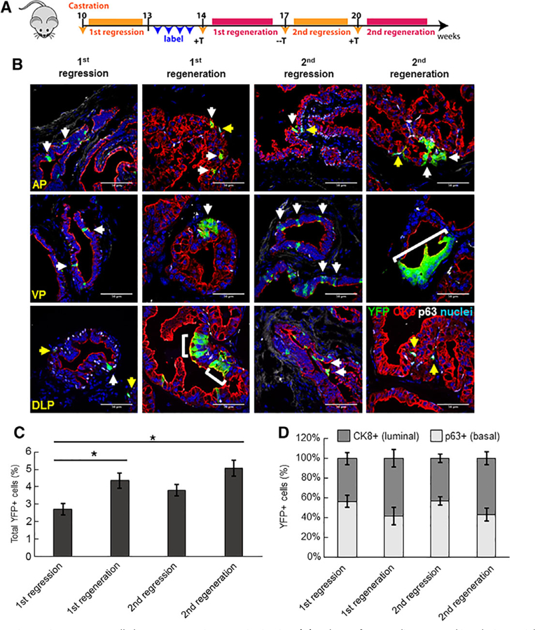
Castration-resistant Sox2+ cells have regenerative capacity in vivo. (A): Scheme for Sox2 lineage marking during serial prostate regression and regeneration. In this approach, Sox2+ cells were labeled for YFP-expression after host castration and prostatic involution. (B): Immunofluorescent staining to assess YFP+/CK8+ luminal cells (white arrows) or YFP+/p63+ basal cells (yellow arrows) after serial prostate regeneration (n = 3 mice per time point). Scale bar: 50 μm. Abbreviations: AP, anterior prostate; VP, ventral prostate; DLP, dorsolateral prostate. (C): Graph showing percentage of total YFP+ cells in intact, castrated or regenerated prostates examined over all lobes. Data represent the mean T SEM; **, p < .01; *, p < .05; Tukey’s Honest Significant Difference test. (D): Graph showing percentage of YFP+ cells coexpressing luminal or basal markers in intact, castrated and regenerated prostates.