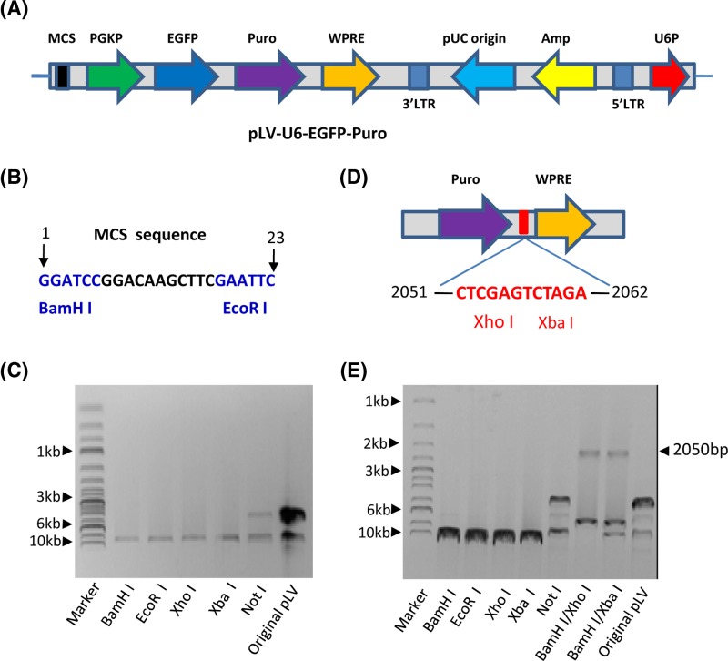
Figure 1. Characterization of the pLV-U6-EGFP-Puro lentiviral vector.
(A) A scheme showing basic elements included in the pLV-U6-EGFP-Puro plasmid. (B) The sequence and location of MCS in the pLV-U6-EGFP-Puro plasmid. The MCS sequence contains only two restriction enzyme sites, BamHI and EcoRI (blue). (C) An agarose gel image showing the result of enzyme digestion of the pLV-U6-EGFP-Puro plasmid. The pLV-U6-EGFP-Puro plasmid was digested with the enzymes as indicated in the Figure, followed by detecting with agarose gel electrophoresis and imaging with ChemiDoc MP imaging system (Bio-Rad). (D) A scheme showing the positions of two restriction enzyme sites, XhoI and XbaI, in the pLV-U6-EGFP-puro plasmid. DNA-sequencing was performed using the pLV-U6-EGFP-Puro plasmid, and the restriction enzyme sites included in the vector were analyzed. (E) Restriction enzyme digestion confirmed that the pLV-U6-EGFP-Puro plasmid contains XhoI and XbaI sites between 2051 and 2063 bp downstream of puromycin resistant gene (Puro). Plasmid DNA was digested using the enzymes as indicated in the Figure; the image for agarose gel electrophoresis was obtained as described in (C).
Abbreviation: original pLV, pLV-U6-EGFP-Puro plasmid.

