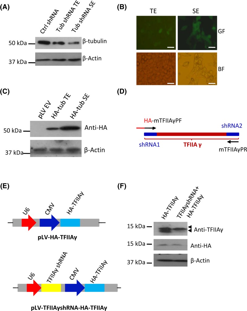
Figure 3. Functional validation of the pLV-U6-CMV-EGFP-Puro plasmid.
(A) The pLV-U6-CMV-EGFP-Puro vector can express shRNA. The pLV-U6-CMV-EGFP-Puro plasmid expressing β-tubulin shRNA was transfected into 293T cells in the presence or absence of packaging vectors. β-tubulin expression was analyzed using both transiently transfected cells and stable cell lines and Western blot. (B) Fluorescence microscopy for the cells that transiently or stably express β-tubulin shRNA. The cells obtained in (A) were imaged under fluorescence microscope. Scale bar in each image represents 50 μm. (C) The pLV-U6-CMV-EGFP-Puro plasmid can express protein. The pLV-U6-CMV-EGFP-Puro plasmid expressing HA-β-tubulin was transfected into 293T cells in the presence or absence of packaging vectors, HA-β-tubulin expression was analyzed using transiently transfected cells and stable cell line and Western blot. (D) A diagram showing TFIIAγ cDNA regions used for TFIIAγ shRNA design and the primers for modification of TFIIAγ cDNA. HA DNA fragment was added to the front of TFIIAγ forward primer to form the HA- mTFIIAγPF fusion primer, the third base of each codon in mTFIIAγPF and mTFIIAγPR was mutated. (E) A diagram showing the plasmids expressing HA-TFIIAγ only (pLV-HA-TFIIAγ) or expressing both TFIIAγ shRNA and HA-TFIIAγ (pLV-TFIIAγshRNA-HA-TFIIAγ). (F) The pLV-TFIIAγshRNA-HA-TFIIAγ plasmid can express TFIIAγ shRNA and HA-TFIIAγ simultaneously. The pLV-TFIIAγshRNA-HA-TFIIAγ and pLV-HA-TFIIAγ plasmids were respectively transfected into 293T cells; after 48 h, expression of TFIIAγ and HA-TFIIAγ was analyzed using transiently transfected cells and Western blot.
Abbreviations: BF, bright field; GF, green fluorescence; SE, stable expression; TE, transient expression.

