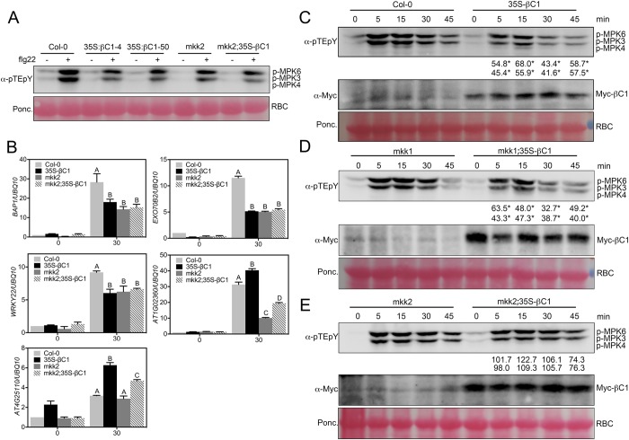
Fig 3. βC1 attenuates flg22 induced MAPK signaling.
(A) Western blot assays show that differential βC1 attenuation of flg22-induced MAPK activation in Col-0 and mkk2 background. (B) qRT-PCR assays show that βC1 inhibition of flg22-induced gene expression in Col-0 and mkk2 background. 10-d seedlings were treated with 1μM flg22 for 30 min. Different letters indicate significant differences among samples (p<0.01, t test). (C) Western blot assays show that βC1 attenuates flg22-induced MAPK activation in vivo. Ten-day-old seedlings were treated with 100 nM flg22 for indicated time windows. MAPK activation was detected by the immunoblot assay with an anti-pTEpY antibody. Myc-βC1 was detected by an anti-Myc antibody and Ponceau S staning of Rubisco (RBC) shows protein loading. (D) Western blot assays show that βC1 attenuation of flg22-induced MAPK activation is largely independent on MKK1 protein. (E) Western blot assays show that βC1 attenuation of flg22-induced MAPK activation is largely dependent on MKK2 protein. Three biological replicates of these experiments were performed. Numbers indicate the average ratio of phosphorylated MPK6 and MPK3 protein in 35S-Myc-βC1 transgenic plants compared wild type at the same flg22 treatment time period for three biological replicates, asterisks represent statistically significant based on Student’s t test at P < 0.05.

