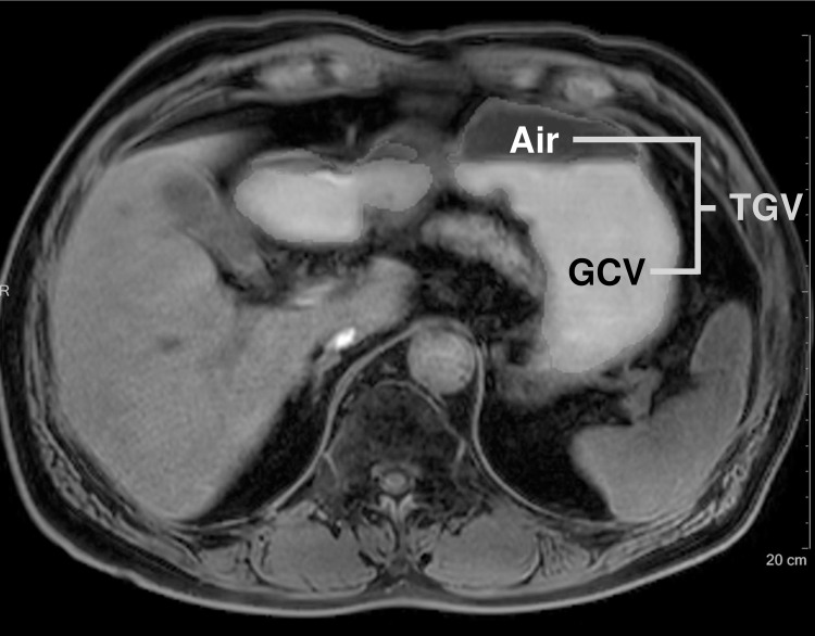
Fig 3. Gastric volume measurement of a 68-year-old man with Parkinson’s disease.
In axial T1-weighted image, regions of interest (ROI) of gastric contents and air were drawn by a radiologist with volume analysis software. The volume of ROI was calculated automatically. TGV, total gastric volume, GCV, gastric content volume.

